6DIO

Structure of class II HMG-CoA reductase from Delftia acidovorans with NAD bound


Experimental Data Snapshot

  • Method: X-RAY DIFFRACTION
  • Resolution: 2.14 Å
  • R-Value Free: 0.239 
  • R-Value Work: 0.191 
  • R-Value Observed: 0.193 

wwPDB Validation   3D Report Full Report


Ligand Structure Quality Assessment 


This is version 1.4 of the entry. See complete history


Literature

New Crystallographic Snapshots of Large Domain Movements in Bacterial 3-Hydroxy-3-methylglutaryl Coenzyme A Reductase.

Ragwan, E.R.Arai, E.Kung, Y.

(2018) Biochemistry 57: 5715-5725

  • DOI: https://doi.org/10.1021/acs.biochem.8b00869
  • Primary Citation of Related Structures:  
    6DIO, 6EEU, 6EEV

  • PubMed Abstract: 

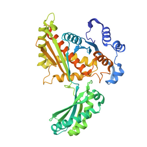
    The enzyme 3-hydroxy-3-methylglutaryl coenzyme A (HMG-CoA) reductase (HMGR) catalyzes the first committed step of the mevalonate pathway, which is used across biology in the biosynthesis of countless metabolites. HMGR consumes 2 equiv of the cofactor NAD(P)H to perform the four-electron reduction of HMG-CoA to mevalonate toward the production of steroids and isoprenoids, the largest class of natural products. Recent structural data have shown that HMGR contains a highly mobile C-terminal domain (CTD) that is believed to adopt many different conformations to permit binding and dissociation of the substrate, cofactors, and products at specific points during the reaction cycle. Here, we have characterized the HMGR from Delftia acidovorans as an NADH-specific enzyme and determined crystal structures of the enzyme in unbound, mevalonate-bound, and NADH- and citrate-bound states. Together, these structures depict ligand binding in both the active site and the cofactor-binding site while illustrating how a conserved helical motif confers NAD(P)H cofactor specificity. Unexpectedly, the NADH-bound structure also reveals a new conformation of the CTD, in which the domain has "flipped" upside-down, while directly binding the cofactor. By capturing these structural snapshots, this work not only expands the known range of HMGR domain movement but also provides valuable insight into the catalytic mechanism of this biologically important enzyme.


  • Organizational Affiliation

    Department of Chemistry , Bryn Mawr College , 101 North Merion Avenue , Bryn Mawr , Pennsylvania 19010 , United States.


Macromolecules
Find similar proteins by:  (by identity cutoff)  |  3D Structure
Entity ID: 1
MoleculeChains Sequence LengthOrganismDetailsImage
3-hydroxy-3-methylglutaryl coenzyme A reductase
A, B, C
429Delftia acidovoransMutation(s): 0 
Gene Names: CHL79_09160
EC: 1.1.1.88
UniProt
Find proteins for A9BQX8 (Delftia acidovorans (strain DSM 14801 / SPH-1))
Explore A9BQX8 
Go to UniProtKB:  A9BQX8
Entity Groups  
Sequence Clusters30% Identity50% Identity70% Identity90% Identity95% Identity100% Identity
UniProt GroupA9BQX8
Sequence Annotations
Expand
  • Reference Sequence
Small Molecules
Ligands 4 Unique
IDChains Name / Formula / InChI Key2D Diagram3D Interactions
NAD (Subject of Investigation/LOI)
Query on NAD

Download Ideal Coordinates CCD File 
D [auth A],
HA [auth C],
O [auth B]
NICOTINAMIDE-ADENINE-DINUCLEOTIDE
C21 H27 N7 O14 P2
BAWFJGJZGIEFAR-NNYOXOHSSA-N
CIT
Query on CIT

Download Ideal Coordinates CCD File 
E [auth A],
IA [auth C],
P [auth B]
CITRIC ACID
C6 H8 O7
KRKNYBCHXYNGOX-UHFFFAOYSA-N
SO4
Query on SO4

Download Ideal Coordinates CCD File 
F [auth A]
G [auth A]
JA [auth C]
KA [auth C]
Q [auth B]
F [auth A],
G [auth A],
JA [auth C],
KA [auth C],
Q [auth B],
R [auth B],
S [auth B],
T [auth B],
U [auth B],
V [auth B]
SULFATE ION
O4 S
QAOWNCQODCNURD-UHFFFAOYSA-L
EDO
Query on EDO

Download Ideal Coordinates CCD File 
AA [auth B]
BA [auth B]
CA [auth B]
DA [auth B]
EA [auth B]
AA [auth B],
BA [auth B],
CA [auth B],
DA [auth B],
EA [auth B],
FA [auth B],
GA [auth B],
H [auth A],
I [auth A],
J [auth A],
K [auth A],
L [auth A],
LA [auth C],
M [auth A],
MA [auth C],
N [auth A],
NA [auth C],
OA [auth C],
PA [auth C],
QA [auth C],
RA [auth C],
SA [auth C],
TA [auth C],
UA [auth C],
W [auth B],
X [auth B],
Y [auth B],
Z [auth B]
1,2-ETHANEDIOL
C2 H6 O2
LYCAIKOWRPUZTN-UHFFFAOYSA-N
Experimental Data & Validation

Experimental Data

  • Method: X-RAY DIFFRACTION
  • Resolution: 2.14 Å
  • R-Value Free: 0.239 
  • R-Value Work: 0.191 
  • R-Value Observed: 0.193 
  • Space Group: P 65 2 2
Unit Cell:
Length ( Å )Angle ( ˚ )
a = 113α = 90
b = 113β = 90
c = 437.48γ = 120
Software Package:
Software NamePurpose
PHENIXrefinement
MOSFLMdata reduction
Aimlessdata scaling
PHASERphasing

Structure Validation

View Full Validation Report


Ligand Structure Quality Assessment 


Entry History & Funding Information

Deposition Data


Funding OrganizationLocationGrant Number
National Institutes of Health/National Institute of General Medical Sciences (NIH/NIGMS)United StatesGM116029

Revision History  (Full details and data files)

  • Version 1.0: 2018-09-26
    Type: Initial release
  • Version 1.1: 2018-10-03
    Changes: Data collection, Database references
  • Version 1.2: 2018-10-17
    Changes: Data collection, Database references
  • Version 1.3: 2020-01-01
    Changes: Author supporting evidence
  • Version 1.4: 2024-03-13
    Changes: Data collection, Database references