6DCD

Mycobacterium marinum cytochrome P450 CYP150A6 in the substrate-free form


Experimental Data Snapshot

  • Method: X-RAY DIFFRACTION
  • Resolution: 1.55 Å
  • R-Value Free: 0.246 
  • R-Value Work: 0.222 
  • R-Value Observed: 0.223 

wwPDB Validation   3D Report Full Report


Ligand Structure Quality Assessment 


This is version 1.3 of the entry. See complete history


Literature

The characterisation of two members of the cytochrome P450 CYP150 family: CYP150A5 and CYP150A6 from Mycobacterium marinum.

Child, S.A.Flint, K.L.Bruning, J.B.Bell, S.G.

(2019) Biochim Biophys Acta Gen Subj 1863: 925-934

  • DOI: https://doi.org/10.1016/j.bbagen.2019.02.016
  • Primary Citation of Related Structures:  
    6DCD

  • PubMed Abstract: 

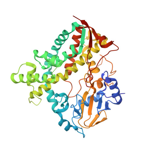
    Actinobacteria, including the Mycobacteria, have a large component of cytochrome P450 family monooxygenases. This includes Mycobacterium tuberculosis, M. ulcerans and M. marinum, and M. vanbaalenii. These enzymes can abstract CH bonds and have important roles in natural product biosynthesis. Two members of the bacterial CYP150 family, CYP150A5 and CYP150A6 from M. marinum, were produced, purified and characterised. The potential substrate ranges of both enzymes were analysed and the monooxygenase activity of CYP150A5 was reconstituted using a physiological electron transfer partner system. CYP150A6 was structurally characterised by X-ray crystallography. CYP150A5 was shown to bind various norisoprenoids and terpenoids. It could regioselectively hydroxylate β-ionol. The X-ray crystal structure of substrate-free CYP150A6 was solved to 1.5 Å. This displayed an open conformation with short F and G helices, an unresolved F-G loop region and exposed active site pocket. The active site residues could be identified and important variations were found among the CYP150A enzymes. Haem-binding azole inhibitors were identified for both enzymes. The structure of CYP150A6 will facilitate the identification of physiological substrates and the design of better inhibitors for members of this P450 family. Based on the observed differences in substrate binding preference and sequence variations among the active site residues, their roles are predicted to be different. Multiple CYP150 family members were found in many bacteria and are prevalent in the Mycobacteria including several human pathogens. Inhibition and structural data are reported here for these enzymes for the first time.


  • Organizational Affiliation

    Department of Chemistry, University of Adelaide, SA 5005, Australia.


Macromolecules
Find similar proteins by:  (by identity cutoff)  |  3D Structure
Entity ID: 1
MoleculeChains Sequence LengthOrganismDetailsImage
Cytochrome P450 150A6 Cyp150A6424Mycobacterium marinum MMutation(s): 0 
Gene Names: cyp150A6MMAR_4694
UniProt
Find proteins for B2HFD6 (Mycobacterium marinum (strain ATCC BAA-535 / M))
Explore B2HFD6 
Go to UniProtKB:  B2HFD6
Entity Groups  
Sequence Clusters30% Identity50% Identity70% Identity90% Identity95% Identity100% Identity
UniProt GroupB2HFD6
Sequence Annotations
Expand
  • Reference Sequence
Small Molecules
Ligands 1 Unique
IDChains Name / Formula / InChI Key2D Diagram3D Interactions
HEM
Query on HEM

Download Ideal Coordinates CCD File 
B [auth A]PROTOPORPHYRIN IX CONTAINING FE
C34 H32 Fe N4 O4
KABFMIBPWCXCRK-RGGAHWMASA-L
Experimental Data & Validation

Experimental Data

  • Method: X-RAY DIFFRACTION
  • Resolution: 1.55 Å
  • R-Value Free: 0.246 
  • R-Value Work: 0.222 
  • R-Value Observed: 0.223 
  • Space Group: P 31 2 1
Unit Cell:
Length ( Å )Angle ( ˚ )
a = 80.57α = 90
b = 80.57β = 90
c = 134.76γ = 120
Software Package:
Software NamePurpose
PHENIXrefinement
MOSFLMdata reduction
Aimlessdata scaling
PHASERphasing
PDB_EXTRACTdata extraction

Structure Validation

View Full Validation Report


Ligand Structure Quality Assessment 


Entry History & Funding Information

Deposition Data


Funding OrganizationLocationGrant Number
Australian Research Council (ARC)AustraliaFT140100355

Revision History  (Full details and data files)

  • Version 1.0: 2019-03-20
    Type: Initial release
  • Version 1.1: 2019-03-27
    Changes: Data collection, Database references
  • Version 1.2: 2020-01-01
    Changes: Author supporting evidence
  • Version 1.3: 2024-03-13
    Changes: Data collection, Database references