


Crystal structure of an organic hydroperoxide resistance protein from Elizabethkingia anophelis with crystallant-derived thiocyanate bound
Mayclin, S.J., Delker, S.L., Horanyi, P.S., Lorimer, D.D., Edwards, T.E.To be published.
Experimental Data Snapshot
wwPDB Validation 3D Report Full Report

Entity ID: 1 | |||||
|---|---|---|---|---|---|
| Molecule | Chains | Sequence Length | Organism | Details | Image |
| Organic hydroperoxide resistance protein | 148 | Elizabethkingia anophelis NUHP1 | Mutation(s): 0 Gene Names: BD94_1921 | 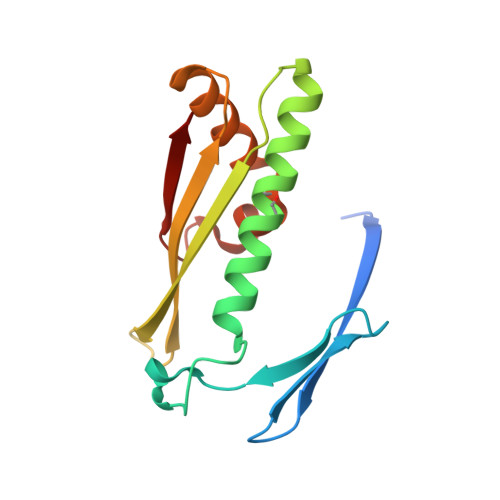 | |
UniProt | |||||
Find proteins for A0A077EDR1 (Elizabethkingia anophelis NUHP1) Explore A0A077EDR1 Go to UniProtKB: A0A077EDR1 | |||||
Entity Groups | |||||
| Sequence Clusters | 30% Identity50% Identity70% Identity90% Identity95% Identity100% Identity | ||||
| UniProt Group | A0A077EDR1 | ||||
Sequence AnnotationsExpand | |||||
| |||||
| Ligands 2 Unique | |||||
|---|---|---|---|---|---|
| ID | Chains | Name / Formula / InChI Key | 2D Diagram | 3D Interactions | |
| EDO Query on EDO | E [auth B], F [auth B] | 1,2-ETHANEDIOL C2 H6 O2 LYCAIKOWRPUZTN-UHFFFAOYSA-N |  | ||
| SCN Query on SCN | C [auth A], D [auth A] | THIOCYANATE ION C N S ZMZDMBWJUHKJPS-UHFFFAOYSA-M |  | ||
| Length ( Å ) | Angle ( ˚ ) |
|---|---|
| a = 47.39 | α = 90 |
| b = 50.03 | β = 102.63 |
| c = 57.98 | γ = 90 |
| Software Name | Purpose |
|---|---|
| PHENIX | refinement |
| XDS | data reduction |
| XSCALE | data scaling |
| MOLREP | phasing |
| PDB_EXTRACT | data extraction |
| Coot | model building |
RCSB PDB (citation) is hosted by
RCSB PDB is a member of the

