6D7J

The Crystal Structure of Parabacteroides merdae Beta-Glucuronidase (GUS) with Glycerol in Active-Site


Experimental Data Snapshot

  • Method: X-RAY DIFFRACTION
  • Resolution: 2.24 Å
  • R-Value Free: 0.200 
  • R-Value Work: 0.165 
  • R-Value Observed: 0.165 

wwPDB Validation   3D Report Full Report


This is version 1.4 of the entry. See complete history


Literature

Active site flexibility revealed in crystal structures of Parabacteroides merdae beta-glucuronidase from the human gut microbiome.

Little, M.S.Ervin, S.M.Walton, W.G.Tripathy, A.Xu, Y.Liu, J.Redinbo, M.R.

(2018) Protein Sci 27: 2010-2022

  • DOI: https://doi.org/10.1002/pro.3507
  • Primary Citation of Related Structures:  
    6D7J, 6DXU

  • PubMed Abstract: 

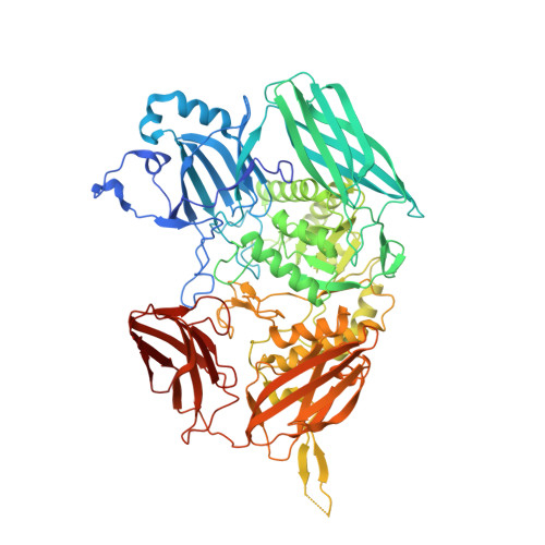
    β-Glucuronidase (GUS) enzymes in the gastrointestinal tract are involved in maintaining mammalian-microbial symbiosis and can play key roles in drug efficacy and toxicity. Parabacteroides merdae GUS was identified as an abundant mini-Loop 2 (mL2) type GUS enzyme in the Human Microbiome Project gut metagenomic database. Here, we report the crystal structure of P. merdae GUS and highlight the differences between this enzyme and extant structures of gut microbial GUS proteins. We find that P. merdae GUS exhibits a distinct tetrameric quaternary structure and that the mL2 motif traces a unique path within the active site, which also includes two arginines distinctive to this GUS. We observe two states of the P. merdae GUS active site; a loop repositions itself by more than 50 Å to place a functionally-relevant residue into the enzyme's catalytic site. Finally, we find that P. merdae GUS is able to bind to homo and heteropolymers of the polysaccharide alginic acid. Together, these data broaden our understanding of the structural and functional diversity in the GUS family of enzymes present in the human gut microbiome and point to specialization as an important feature of microbial GUS orthologs.


  • Organizational Affiliation

    Department of Chemistry, University of North Carolina, Chapel Hill, North Carolina, 27599-3290.


Macromolecules
Find similar proteins by:  (by identity cutoff)  |  3D Structure
Entity ID: 1
MoleculeChains Sequence LengthOrganismDetailsImage
Beta-Glucuronidase
A, B, C, D
830Parabacteroides merdae CL03T12C32Mutation(s): 0 
Gene Names: HMPREF1060_00403
UniProt
Find proteins for K5ZWV5 (Parabacteroides merdae CL03T12C32)
Explore K5ZWV5 
Go to UniProtKB:  K5ZWV5
Entity Groups  
Sequence Clusters30% Identity50% Identity70% Identity90% Identity95% Identity100% Identity
UniProt GroupK5ZWV5
Sequence Annotations
Expand
  • Reference Sequence
Small Molecules
Ligands 4 Unique
IDChains Name / Formula / InChI Key2D Diagram3D Interactions
PRO
Query on PRO

Download Ideal Coordinates CCD File 
NB [auth D],
R [auth A]
PROLINE
C5 H9 N O2
ONIBWKKTOPOVIA-BYPYZUCNSA-N
GOL
Query on GOL

Download Ideal Coordinates CCD File 
AA [auth B]
AB [auth C]
BA [auth B]
BB [auth C]
CA [auth B]
AA [auth B],
AB [auth C],
BA [auth B],
BB [auth C],
CA [auth B],
CB [auth C],
DA [auth B],
EA [auth B],
F [auth A],
FA [auth B],
FB [auth D],
G [auth A],
GA [auth B],
GB [auth D],
H [auth A],
HA [auth B],
HB [auth D],
I [auth A],
IA [auth B],
IB [auth D],
J [auth A],
JA [auth B],
JB [auth D],
K [auth A],
KB [auth D],
L [auth A],
LB [auth D],
M [auth A],
MA [auth C],
MB [auth D],
N [auth A],
NA [auth C],
O [auth A],
OA [auth C],
P [auth A],
PA [auth C],
Q [auth A],
QA [auth C],
RA [auth C],
SA [auth C],
TA [auth C],
U [auth B],
UA [auth C],
V [auth B],
VA [auth C],
W [auth B],
WA [auth C],
X [auth B],
XA [auth C],
Y [auth B],
YA [auth C],
Z [auth B],
ZA [auth C]
GLYCEROL
C3 H8 O3
PEDCQBHIVMGVHV-UHFFFAOYSA-N
K
Query on K

Download Ideal Coordinates CCD File 
E [auth A],
EB [auth D],
LA [auth C],
T [auth B]
POTASSIUM ION
K
NPYPAHLBTDXSSS-UHFFFAOYSA-N
NA
Query on NA

Download Ideal Coordinates CCD File 
DB [auth C],
KA [auth B],
OB [auth D],
S [auth A]
SODIUM ION
Na
FKNQFGJONOIPTF-UHFFFAOYSA-N
Experimental Data & Validation

Experimental Data

  • Method: X-RAY DIFFRACTION
  • Resolution: 2.24 Å
  • R-Value Free: 0.200 
  • R-Value Work: 0.165 
  • R-Value Observed: 0.165 
  • Space Group: P 1 21 1
Unit Cell:
Length ( Å )Angle ( ˚ )
a = 92.92α = 90
b = 171.968β = 107.84
c = 125.322γ = 90
Software Package:
Software NamePurpose
PHENIXphasing
PHENIXrefinement
HKL-2000data scaling
PHENIXmodel building
HKL-2000data reduction

Structure Validation

View Full Validation Report


Entry History & Funding Information

Deposition Data


Funding OrganizationLocationGrant Number
National Institutes of Health/National Human Genome Research Institute (NIH/NHGRI)United StatesCA098468
National Institutes of Health/National Human Genome Research Institute (NIH/NHGRI)United StatesCA207416

Revision History  (Full details and data files)

  • Version 1.0: 2019-05-01
    Type: Initial release
  • Version 1.1: 2019-12-18
    Changes: Author supporting evidence
  • Version 1.2: 2020-05-13
    Changes: Database references
  • Version 1.3: 2020-05-20
    Changes: Database references
  • Version 1.4: 2024-03-13
    Changes: Data collection, Database references, Derived calculations, Refinement description