Structural basis of Toxoplasma gondii perforin-like protein 1 membrane interaction and activity during egress.
Guerra, A.J., Zhang, O., Bahr, C.M.E., Huynh, M.H., DelProposto, J., Brown, W.C., Wawrzak, Z., Koropatkin, N.M., Carruthers, V.B.(2018) PLoS Pathog 14: e1007476-e1007476
- PubMed: 30513119
- DOI: https://doi.org/10.1371/journal.ppat.1007476
- Primary Citation of Related Structures:
6D7A - PubMed Abstract:
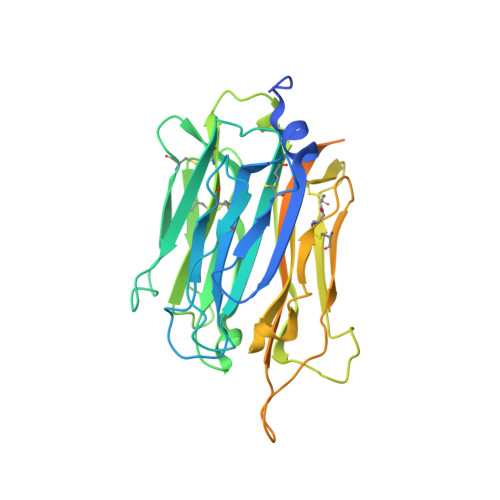
Intracellular pathogens must egress from the host cell to continue their infectious cycle. Apicomplexans are a phylum of intracellular protozoans that have evolved members of the membrane attack complex and perforin (MACPF) family of pore forming proteins to disrupt cellular membranes for traversing cells during tissue migration or egress from a replicative vacuole following intracellular reproduction. Previous work showed that the apicomplexan Toxoplasma gondii secretes a perforin-like protein (TgPLP1) that contains a C-terminal Domain (CTD) which is necessary for efficient parasite egress. However, the structural basis for CTD membrane binding and egress competency remained unknown. Here, we present evidence that TgPLP1 CTD prefers binding lipids that are abundant in the inner leaflet of the lipid bilayer. Additionally, solving the high-resolution crystal structure of the TgPLP1 APCβ domain within the CTD reveals an unusual double-layered β-prism fold that resembles only one other protein of known structure. Three direct repeat sequences comprise subdomains, with each constituting a wall of the β-prism fold. One subdomain features a protruding hydrophobic loop with an exposed tryptophan at its tip. Spectrophotometric measurements of intrinsic tryptophan fluorescence are consistent with insertion of the hydrophobic loop into a target membrane. Using CRISPR/Cas9 gene editing we show that parasite strains bearing mutations in the hydrophobic loop, including alanine substitution of the tip tryptophan, are equally deficient in egress as a strain lacking TgPLP1 altogether. Taken together our findings suggest a crucial role for the hydrophobic loop in anchoring TgPLP1 to the membrane to support its cytolytic activity and egress function.
Organizational Affiliation:
Department of Microbiology and Immunology, University of Michigan, Ann Arbor, MI, United States of America.