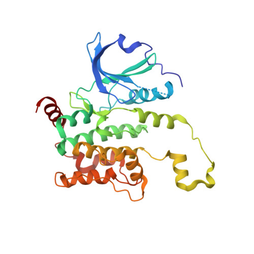
6CQD

Crystal structure of HPK1 in complex with ATP analogue (AMPPNP)


Experimental Data Snapshot

  • Method: X-RAY DIFFRACTION
  • Resolution: 2.12 Å
  • R-Value Free: 0.250 
  • R-Value Work: 0.203 
  • R-Value Observed: 0.206 

wwPDB Validation   3D Report Full Report


Ligand Structure Quality Assessment 


This is version 1.2 of the entry. See complete history


Literature

Hematopoietic Progenitor Kinase-1 Structure in a Domain-Swapped Dimer.

Wu, P.Sneeringer, C.J.Pitts, K.E.Day, E.S.Chan, B.K.Wei, B.Lehoux, I.Mortara, K.Li, H.Wu, J.Franke, Y.Moffat, J.G.Grogan, J.L.Heffron, T.P.Wang, W.

(2019) Structure 27: 125-133.e4

  • DOI: https://doi.org/10.1016/j.str.2018.10.025
  • Primary Citation of Related Structures:  
    6CQD, 6CQE, 6CQF

  • PubMed Abstract: 

    Enhancement of antigen-specific T cell immunity has shown significant therapeutic benefit in infectious diseases and cancer. Hematopoietic progenitor kinase-1 (HPK1) is a negative-feedback regulator of T cell receptor signaling, which dampens T cell proliferation and effector function. A recent report showed that a catalytic dead mutant of HPK1 phenocopies augmented T cell responses observed in HPK1-knockout mice, indicating that kinase activity is critical for function. We evaluated active and inactive mutants and determined crystal structures of HPK1 kinase domain (HPK1-KD) in apo and ligand bound forms. In all structures HPK1-KD displays a rare domain-swapped dimer, in which the activation segment comprises a well-conserved dimer interface. Biophysical measurements show formation of dimer in solution. The activation segment adopts an α-helical structure which exhibits distinct orientations in active and inactive states. This face-to-face configuration suggests that the domain-swapped dimer may possess alternative selectivity for certain substrates of HPK1 under relevant cellular context.


  • Organizational Affiliation

    Department of Structural Biology, Genentech, South San Francisco, CA 94080, USA.


Macromolecules
Find similar proteins by:  (by identity cutoff)  |  3D Structure
Entity ID: 1
MoleculeChains Sequence LengthOrganismDetailsImage
Mitogen-activated protein kinase kinase kinase kinase 1
A, B
297Homo sapiensMutation(s): 0 
Gene Names: MAP4K1HPK1
EC: 2.7.11.1
UniProt & NIH Common Fund Data Resources
Find proteins for Q92918 (Homo sapiens)
Explore Q92918 
Go to UniProtKB:  Q92918
PHAROS:  Q92918
GTEx:  ENSG00000104814 
Entity Groups  
Sequence Clusters30% Identity50% Identity70% Identity90% Identity95% Identity100% Identity
UniProt GroupQ92918
Sequence Annotations
Expand
  • Reference Sequence
Experimental Data & Validation

Experimental Data

  • Method: X-RAY DIFFRACTION
  • Resolution: 2.12 Å
  • R-Value Free: 0.250 
  • R-Value Work: 0.203 
  • R-Value Observed: 0.206 
  • Space Group: C 1 2 1
Unit Cell:
Length ( Å )Angle ( ˚ )
a = 146.886α = 90
b = 51.882β = 131.03
c = 107.5γ = 90
Software Package:
Software NamePurpose
PHENIXrefinement
XDSdata reduction
Aimlessdata scaling
PHASERphasing

Structure Validation

View Full Validation Report


Ligand Structure Quality Assessment 


Entry History 

Deposition Data

Revision History  (Full details and data files)

  • Version 1.0: 2018-12-19
    Type: Initial release
  • Version 1.1: 2019-01-16
    Changes: Data collection, Database references
  • Version 1.2: 2024-03-13
    Changes: Data collection, Database references, Derived calculations