


Crystal Structure of Biotin Acetyl Coenzyme A Carboxylase Synthetase from Helicobacter pylori with bound Biotinylated ATP
Dranow, D.M., Horanyi, P.S., Lorimer, D.D., Edwards, T.E.To be published.
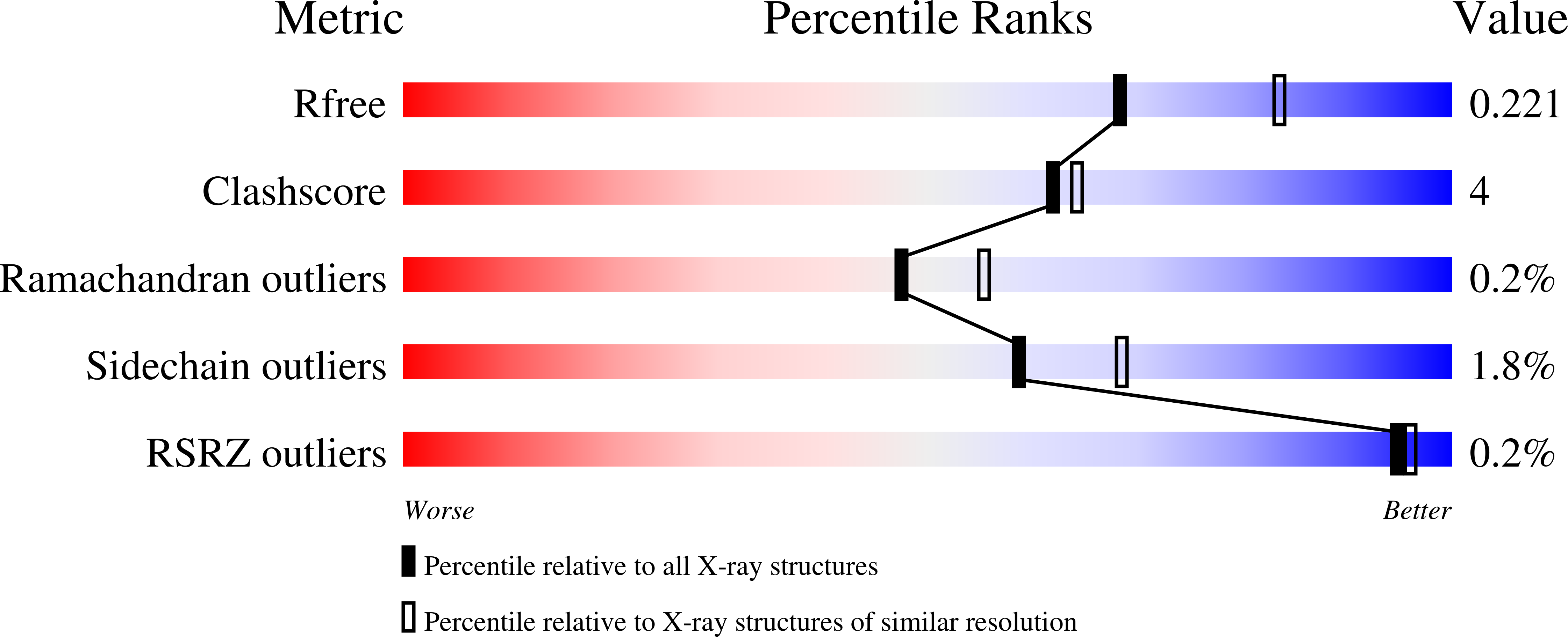
Experimental Data Snapshot
Starting Model: experimental
View more details
Entity ID: 1 | |||||
|---|---|---|---|---|---|
| Molecule | Chains | Sequence Length | Organism | Details | Image |
| Biotin acetyl coenzyme A carboxylase synthetase | 220 | Helicobacter pylori G27 | Mutation(s): 0 Gene Names: HPG27_1085 | 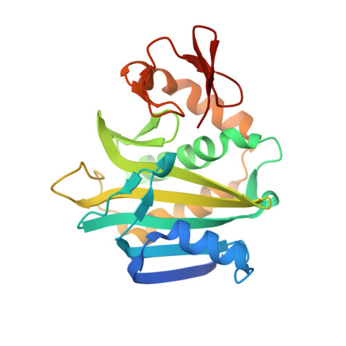 | |
UniProt | |||||
Find proteins for B5Z8D8 (Helicobacter pylori (strain G27)) Explore B5Z8D8 Go to UniProtKB: B5Z8D8 | |||||
Entity Groups | |||||
| Sequence Clusters | 30% Identity50% Identity70% Identity90% Identity95% Identity100% Identity | ||||
| UniProt Group | B5Z8D8 | ||||
Sequence AnnotationsExpand | |||||
| |||||
| Ligands 3 Unique | |||||
|---|---|---|---|---|---|
| ID | Chains | Name / Formula / InChI Key | 2D Diagram | 3D Interactions | |
| F5D (Subject of Investigation/LOI) Query on F5D | C [auth A], F [auth B] | 5'-O-[(S)-({5-[(2R,3aS,4S,6aR)-2-hydroxyhexahydro-1H-thieno[3,4-d]imidazol-4-yl]pentanoyl}oxy){[(S)-hydroxy(phosphonooxy)phosphoryl]oxy}phosphoryl]adenosine C20 H32 N7 O15 P3 S KMVPKOZRCVFEFI-NSWRTZSASA-N |  | ||
| SO4 Query on SO4 | D [auth A], G [auth B], H [auth B], I [auth B] | SULFATE ION O4 S QAOWNCQODCNURD-UHFFFAOYSA-L |  | ||
| EDO Query on EDO | E [auth A], J [auth B], K [auth B], L [auth B] | 1,2-ETHANEDIOL C2 H6 O2 LYCAIKOWRPUZTN-UHFFFAOYSA-N |  | ||
| Length ( Å ) | Angle ( ˚ ) |
|---|---|
| a = 52.89 | α = 122.14 |
| b = 57.08 | β = 94.73 |
| c = 57.03 | γ = 107.85 |
| Software Name | Purpose |
|---|---|
| XDS | data reduction |
| XSCALE | data scaling |
| PHENIX | refinement |
| PDB_EXTRACT | data extraction |
| MR-Rosetta | phasing |
RCSB PDB (citation) is hosted by
RCSB PDB is a member of the

