6C5Y

Crystal structure of thaumatin from microcrystals


Experimental Data Snapshot

  • Method: X-RAY DIFFRACTION
  • Resolution: 2.50 Å
  • R-Value Free: 0.220 
  • R-Value Work: 0.182 
  • R-Value Observed: 0.184 

wwPDB Validation   3D Report Full Report


This is version 1.3 of the entry. See complete history


Literature

Sample manipulation and data assembly for robust microcrystal synchrotron crystallography.

Guo, G.Fuchs, M.R.Shi, W.Skinner, J.Berman, E.Ogata, C.M.Hendrickson, W.A.McSweeney, S.Liu, Q.

(2018) IUCrJ 5: 238-246

  • DOI: https://doi.org/10.1107/S2052252518005389
  • Primary Citation of Related Structures:  
    6C5Y

  • PubMed Abstract: 

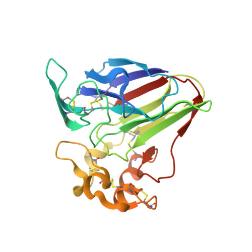
    With the recent developments in microcrystal handling, synchrotron microdiffraction beamline instrumentation and data analysis, microcrystal crystallo-graphy with crystal sizes of less than 10 µm is appealing at synchrotrons. However, challenges remain in sample manipulation and data assembly for robust microcrystal synchrotron crystallography. Here, the development of micro-sized polyimide well-mounts for the manipulation of microcrystals of a few micrometres in size and the implementation of a robust data-analysis method for the assembly of rotational microdiffraction data sets from many microcrystals are described. The method demonstrates that microcrystals may be routinely utilized for the acquisition and assembly of complete data sets from synchrotron microdiffraction beamlines.


  • Organizational Affiliation

    Photon Science Directorate, NSLS-II, Brookhaven National Laboratory, Upton, NY 11973, USA.


Macromolecules
Find similar proteins by:  (by identity cutoff)  |  3D Structure
Entity ID: 1
MoleculeChains Sequence LengthOrganismDetailsImage
Thaumatin-1207Thaumatococcus danielliiMutation(s): 0 
UniProt
Find proteins for P02883 (Thaumatococcus daniellii)
Explore P02883 
Go to UniProtKB:  P02883
Entity Groups  
Sequence Clusters30% Identity50% Identity70% Identity90% Identity95% Identity100% Identity
UniProt GroupP02883
Sequence Annotations
Expand
  • Reference Sequence
Experimental Data & Validation

Experimental Data

  • Method: X-RAY DIFFRACTION
  • Resolution: 2.50 Å
  • R-Value Free: 0.220 
  • R-Value Work: 0.182 
  • R-Value Observed: 0.184 
  • Space Group: P 41 21 2
Unit Cell:
Length ( Å )Angle ( ˚ )
a = 57.576α = 90
b = 57.576β = 90
c = 150.477γ = 90
Software Package:
Software NamePurpose
PHENIXrefinement
DIALSdata reduction
Aimlessdata scaling
PHASERphasing

Structure Validation

View Full Validation Report


Entry History & Funding Information

Deposition Data


Funding OrganizationLocationGrant Number
National Institutes of Health/National Institute of General Medical Sciences (NIH/NIGMS)United StatesGM107462

Revision History  (Full details and data files)

  • Version 1.0: 2018-05-30
    Type: Initial release
  • Version 1.1: 2019-02-20
    Changes: Author supporting evidence, Data collection
  • Version 1.2: 2020-01-01
    Changes: Author supporting evidence
  • Version 1.3: 2023-10-04
    Changes: Data collection, Database references, Refinement description