6B9I

The crystal structure of the Staphylococcus aureus Fatty acid Kinase (Fak) B1 protein loaded with 14-Methylhexadecanoic Acid (Anteiso C17:0) to 1.93 Angstrom resolution


Experimental Data Snapshot

  • Method: X-RAY DIFFRACTION
  • Resolution: 1.93 Å
  • R-Value Free: 0.206 
  • R-Value Work: 0.166 
  • R-Value Observed: 0.168 

wwPDB Validation   3D Report Full Report


Ligand Structure Quality Assessment 


This is version 1.4 of the entry. See complete history


Literature

Acyl-chain selectivity and physiological roles ofStaphylococcus aureusfatty acid-binding proteins.

Cuypers, M.G.Subramanian, C.Gullett, J.M.Frank, M.W.White, S.W.Rock, C.O.

(2019) J Biol Chem 294: 38-49

  • DOI: https://doi.org/10.1074/jbc.RA118.006160
  • Primary Citation of Related Structures:  
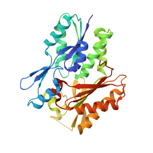
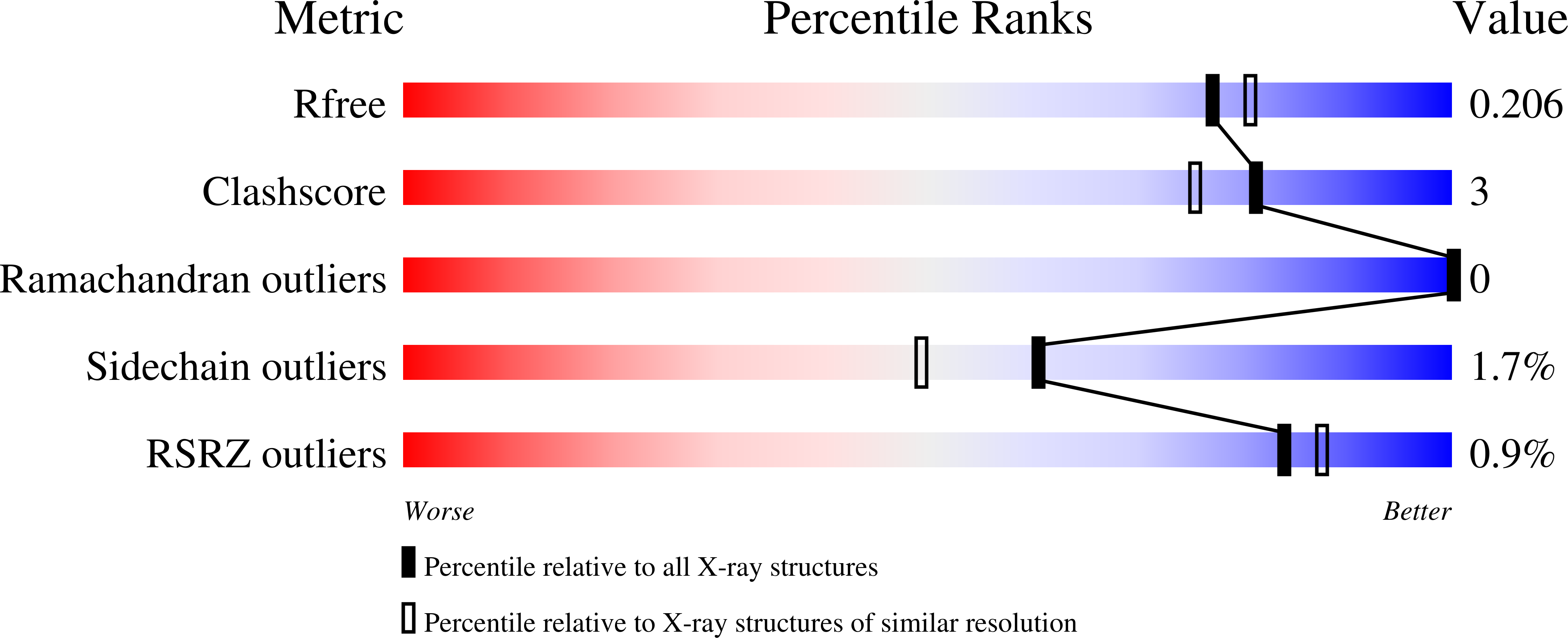
    5WOO, 6ALW, 6B9I

  • PubMed Abstract: 

    Fatty acid (FA) kinase produces acyl-phosphate for the synthesis of membrane phospholipids in Gram-positive bacterial pathogens. FA kinase consists of a kinase protein (FakA) that phosphorylates an FA substrate bound to a second module, an FA-binding protein (FakB). Staphylococcus aureus expresses two distinct, but related, FakBs with different FA selectivities. Here, we report the structures of FakB1 bound to four saturated FAs at 1.6-1.93 Å resolution. We observed that the different FA structures are accommodated within a slightly curved hydrophobic cavity whose length is governed by the conformation of an isoleucine side chain at the end of the tunnel. The hydrophobic tunnel in FakB1 prevents the binding of cis -unsaturated FAs, which are instead accommodated by the kinked tunnel within the FakB2 protein. The differences in the FakB interiors are not propagated to the proteins' surfaces, preserving the protein-protein interactions with their three common partners, FakA, PlsX, and PlsY. Using cellular thermal shift analyses, we found that FakB1 binds FA in vivo , whereas a significant proportion of FakB2 does not. Incorporation of exogenous FA into phospholipid in Δ fakB1 and Δ fakB2 S. aureus knockout strains revealed that FakB1 does not efficiently activate unsaturated FAs. FakB2 preferred unsaturated FAs, but also allowed the incorporation of saturated FAs. These results are consistent with a model in which FakB1 primarily functions in the recycling of the saturated FAs produced by S. aureus metabolism, whereas FakB2 activates host-derived oleate, which S. aureus does not produce but is abundant at infection sites.


  • Organizational Affiliation

    Department of Structural Biology, St. Jude Children's Research Hospital, Memphis, Tennessee 38105.


Macromolecules
Find similar proteins by:  (by identity cutoff)  |  3D Structure
Entity ID: 1
MoleculeChains Sequence LengthOrganismDetailsImage
Fatty acid Kinase (Fak) B1
A, B
288Staphylococcus aureusMutation(s): 0 
Gene Names: AYM28_04055AYM37_04055ERS072738_00223ERS072840_01626ERS074020_00218HMPREF3211_01094
UniProt
Find proteins for X5EH37 (Staphylococcus aureus)
Explore X5EH37 
Go to UniProtKB:  X5EH37
Entity Groups  
Sequence Clusters30% Identity50% Identity70% Identity90% Identity95% Identity100% Identity
UniProt GroupX5EH37
Sequence Annotations
Expand
  • Reference Sequence
Small Molecules
Ligands 3 Unique
IDChains Name / Formula / InChI Key2D Diagram3D Interactions
D0G
Query on D0G

Download Ideal Coordinates CCD File 
C [auth A],
K [auth B]
(14S)-14-methylhexadecanoic acid
C17 H34 O2
FXUKWLSZZHVEJD-INIZCTEOSA-N
GOL
Query on GOL

Download Ideal Coordinates CCD File 
D [auth A]
E [auth A]
F [auth A]
G [auth A]
H [auth A]
D [auth A],
E [auth A],
F [auth A],
G [auth A],
H [auth A],
I [auth A],
J [auth A],
L [auth B],
M [auth B],
N [auth B],
O [auth B]
GLYCEROL
C3 H8 O3
PEDCQBHIVMGVHV-UHFFFAOYSA-N
NA
Query on NA

Download Ideal Coordinates CCD File 
P [auth B],
Q [auth B]
SODIUM ION
Na
FKNQFGJONOIPTF-UHFFFAOYSA-N
Experimental Data & Validation

Experimental Data

  • Method: X-RAY DIFFRACTION
  • Resolution: 1.93 Å
  • R-Value Free: 0.206 
  • R-Value Work: 0.166 
  • R-Value Observed: 0.168 
  • Space Group: P 1
  • Diffraction Data: https://doi.org/10.18430/m36b9i
Unit Cell:
Length ( Å )Angle ( ˚ )
a = 32.991α = 104.71
b = 54.17β = 90
c = 83.901γ = 107.73
Software Package:
Software NamePurpose
PHENIXrefinement
XDSdata reduction
Aimlessdata scaling
MOLREPphasing

Structure Validation

View Full Validation Report


Ligand Structure Quality Assessment 


Entry History 

Deposition Data

Revision History  (Full details and data files)

  • Version 1.0: 2018-10-10
    Type: Initial release
  • Version 1.1: 2018-11-21
    Changes: Data collection, Database references, Structure summary
  • Version 1.2: 2018-11-28
    Changes: Data collection, Database references
  • Version 1.3: 2019-01-16
    Changes: Data collection, Database references
  • Version 1.4: 2023-10-04
    Changes: Data collection, Database references, Refinement description