6AW3

Crystal structure of the HopQ-CEACAM3 L44Q complex


Experimental Data Snapshot

  • Method: X-RAY DIFFRACTION
  • Resolution: 2.66 Å
  • R-Value Free: 0.297 
  • R-Value Work: 0.242 
  • R-Value Observed: 0.245 

wwPDB Validation   3D Report Full Report


This is version 1.1 of the entry. See complete history


Literature

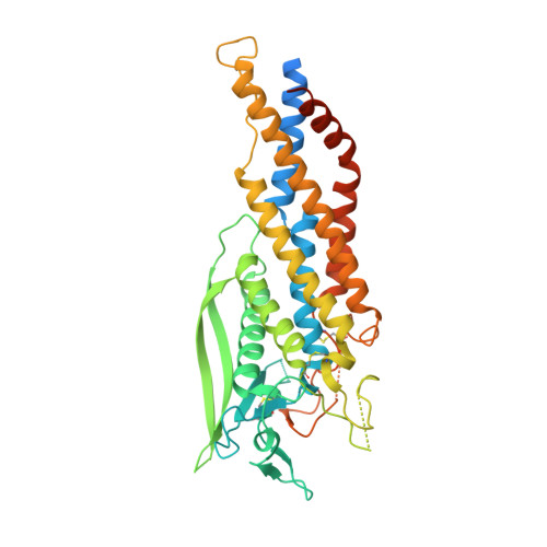
TheHelicobacter pyloriadhesin protein HopQ exploits the dimer interface of human CEACAMs to facilitate translocation of the oncoprotein CagA.

Bonsor, D.A.Zhao, Q.Schmidinger, B.Weiss, E.Wang, J.Deredge, D.Beadenkopf, R.Dow, B.Fischer, W.Beckett, D.Wintrode, P.L.Haas, R.Sundberg, E.J.

(2018) EMBO J 37

  • DOI: https://doi.org/10.15252/embj.201798664
  • Primary Citation of Related Structures:  
    6AVZ, 6AW0, 6AW1, 6AW2, 6AW3

  • PubMed Abstract: 

    Helicobacter pylori infects half of the world's population, and strains that encode the cag type IV secretion system for injection of the oncoprotein CagA into host gastric epithelial cells are associated with elevated levels of cancer. CagA translocation into host cells is dependent on interactions between the H. pylori adhesin protein HopQ and human CEACAMs. Here, we present high-resolution structures of several HopQ-CEACAM complexes and CEACAMs in their monomeric and dimeric forms establishing that HopQ uses a coupled folding and binding mechanism to engage the canonical CEACAM dimerization interface for CEACAM recognition. By combining mutagenesis with biophysical and functional analyses, we show that the modes of CEACAM recognition by HopQ and CEACAMs themselves are starkly different. Our data describe precise molecular mechanisms by which microbes exploit host CEACAMs for infection and enable future development of novel oncoprotein translocation inhibitors and H. pylori -specific antimicrobial agents.


  • Organizational Affiliation

    Institute of Human Virology, University of Maryland School of Medicine, University of Maryland, Baltimore, MD, USA.


Macromolecules
Find similar proteins by:  (by identity cutoff)  |  3D Structure
Entity ID: 1
MoleculeChains Sequence LengthOrganismDetailsImage
HopQA [auth B]439Helicobacter pyloriMutation(s): 0 
Gene Names: hopQ
UniProt
Find proteins for H6A3H4 (Helicobacter pylori)
Explore H6A3H4 
Go to UniProtKB:  H6A3H4
Entity Groups  
Sequence Clusters30% Identity50% Identity70% Identity90% Identity95% Identity100% Identity
UniProt GroupH6A3H4
Sequence Annotations
Expand
  • Reference Sequence
Find similar proteins by:  (by identity cutoff)  |  3D Structure
Entity ID: 2
MoleculeChains Sequence LengthOrganismDetailsImage
Carcinoembryonic antigen-related cell adhesion molecule 3B [auth A]109Homo sapiensMutation(s): 1 
Gene Names: CEACAM3CD66DCGM1
UniProt & NIH Common Fund Data Resources
Find proteins for P40198 (Homo sapiens)
Explore P40198 
Go to UniProtKB:  P40198
PHAROS:  P40198
GTEx:  ENSG00000170956 
Entity Groups  
Sequence Clusters30% Identity50% Identity70% Identity90% Identity95% Identity100% Identity
UniProt GroupP40198
Sequence Annotations
Expand
  • Reference Sequence
Experimental Data & Validation

Experimental Data

  • Method: X-RAY DIFFRACTION
  • Resolution: 2.66 Å
  • R-Value Free: 0.297 
  • R-Value Work: 0.242 
  • R-Value Observed: 0.245 
  • Space Group: P 2 2 21
Unit Cell:
Length ( Å )Angle ( ˚ )
a = 38.83α = 90
b = 102.594β = 90
c = 112.65γ = 90
Software Package:
Software NamePurpose
REFMACrefinement
XDSdata reduction
Aimlessdata scaling
MOLREPphasing

Structure Validation

View Full Validation Report


Entry History 

Deposition Data

Revision History  (Full details and data files)

  • Version 1.0: 2018-05-16
    Type: Initial release
  • Version 1.1: 2018-07-11
    Changes: Data collection, Database references