6AQQ

Crystal structure of Staphylococcus aureus biotin protein ligase in complex with inhibitor


Experimental Data Snapshot

  • Method: X-RAY DIFFRACTION
  • Resolution: 2.71 Å
  • R-Value Free: 0.244 
  • R-Value Work: 0.218 
  • R-Value Observed: 0.219 

wwPDB Validation   3D Report Full Report


Ligand Structure Quality Assessment 


This is version 1.4 of the entry. See complete history


Literature

Halogenation of Biotin Protein Ligase Inhibitors Improves Whole Cell Activity against Staphylococcus aureus.

Paparella, A.S.Lee, K.J.Hayes, A.J.Feng, J.Feng, Z.Cini, D.Deshmukh, S.Booker, G.W.Wilce, M.C.J.Polyak, S.W.Abell, A.D.

(2018) ACS Infect Dis 4: 175-184

  • DOI: https://doi.org/10.1021/acsinfecdis.7b00134
  • Primary Citation of Related Structures:  
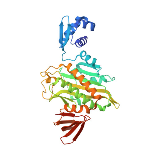
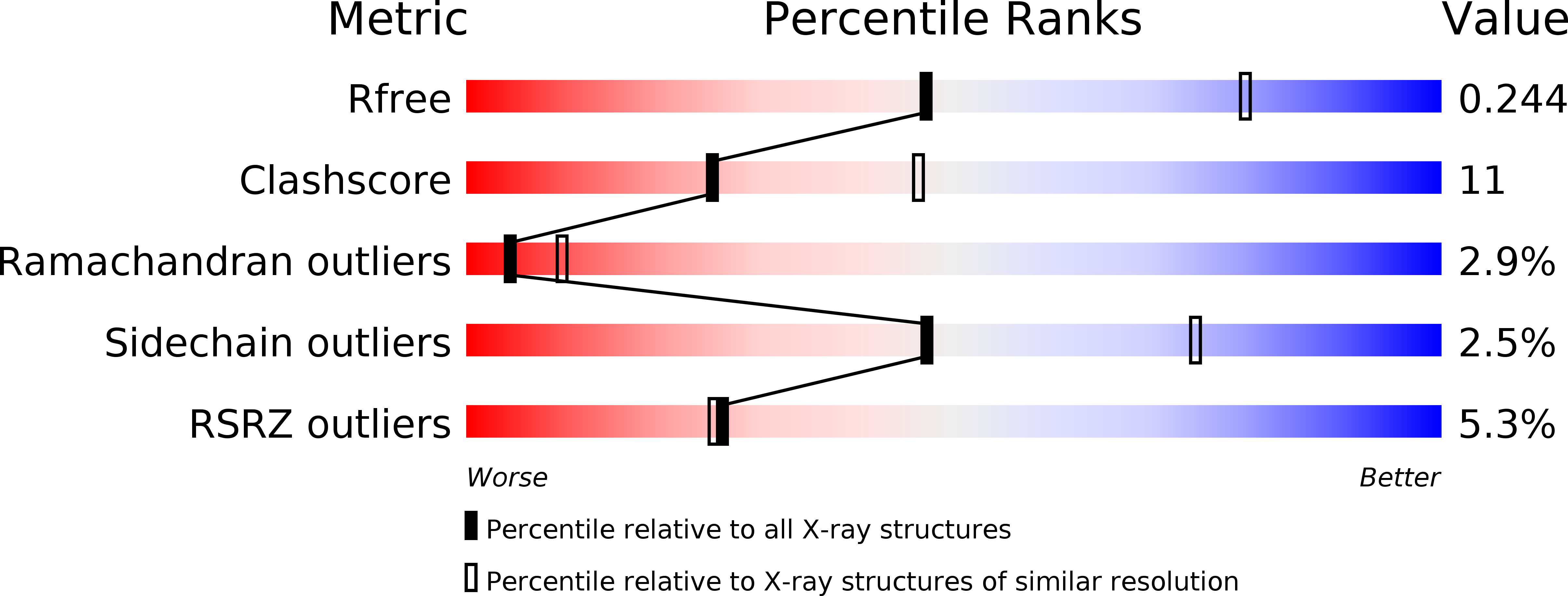
    6APW, 6AQQ, 8ENI

  • PubMed Abstract: 

    We report the synthesis and evaluation of 5-halogenated-1,2,3-triazoles as inhibitors of biotin protein ligase from Staphylococcus aureus. The halogenated compounds exhibit significantly improved antibacterial activity over their nonhalogenated counterparts. Importantly, the 5-fluoro-1,2,3-triazole compound 4c displays antibacterial activity against S. aureus ATCC49775 with a minimum inhibitory concentration (MIC) of 8 μg/mL.


  • Organizational Affiliation

    Department of Molecular and Cellular Biology, University of Adelaide , North Tce, Adelaide, South Australia 5005, Australia.


Macromolecules
Find similar proteins by:  (by identity cutoff)  |  3D Structure
Entity ID: 1
MoleculeChains Sequence LengthOrganismDetailsImage
Bifunctional ligase/repressor BirA329Staphylococcus aureusMutation(s): 0 
Gene Names: birABO217_2143SAMEA2236422_00088SAMEA2236459_00088SAMEA2384800_00088SAMEA2384856_00382SAMEA2445640_00088
EC: 6.3.4.15
Entity Groups  
Sequence Clusters30% Identity50% Identity70% Identity90% Identity95% Identity100% Identity
Sequence Annotations
Expand
  • Reference Sequence
Small Molecules
Ligands 1 Unique
IDChains Name / Formula / InChI Key2D Diagram3D Interactions
BVY
Query on BVY

Download Ideal Coordinates CCD File 
B [auth A](3aS,4S,6aR)-4-(5-{1-[(3-fluorophenyl)methyl]-1H-1,2,3-triazol-4-yl}pentyl)tetrahydro-1H-thieno[3,4-d]imidazol-2(3H)-one
C19 H24 F N5 O S
RJRZSEAZACQQJO-BZSNNMDCSA-N
Experimental Data & Validation

Experimental Data

  • Method: X-RAY DIFFRACTION
  • Resolution: 2.71 Å
  • R-Value Free: 0.244 
  • R-Value Work: 0.218 
  • R-Value Observed: 0.219 
  • Space Group: P 42 21 2
Unit Cell:
Length ( Å )Angle ( ˚ )
a = 93.6α = 90
b = 93.6β = 90
c = 130.7γ = 90
Software Package:
Software NamePurpose
PHENIXrefinement
XDSdata reduction
Aimlessdata scaling
MOLREPphasing

Structure Validation

View Full Validation Report


Ligand Structure Quality Assessment 


Entry History & Funding Information

Deposition Data


Funding OrganizationLocationGrant Number
National Health and Medical Research Council (NHMRC, Australia)AustraliaAPP1068885

Revision History  (Full details and data files)

  • Version 1.0: 2018-02-07
    Type: Initial release
  • Version 1.1: 2018-02-14
    Changes: Author supporting evidence
  • Version 1.2: 2018-02-21
    Changes: Database references
  • Version 1.3: 2020-01-08
    Changes: Author supporting evidence
  • Version 1.4: 2024-03-13
    Changes: Data collection, Database references