6AQJ

Crystal structures of Staphylococcus aureus ketol-acid reductoisomerase in complex with two transition state analogs that have biocidal activity.


Experimental Data Snapshot

  • Method: X-RAY DIFFRACTION
  • Resolution: 1.37 Å
  • R-Value Free: 0.172 
  • R-Value Work: 0.154 
  • R-Value Observed: 0.154 

wwPDB Validation   3D Report Full Report


Ligand Structure Quality Assessment 


This is version 1.3 of the entry. See complete history


Literature

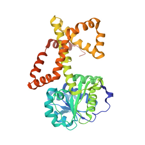
Crystal Structures of Staphylococcus aureus Ketol-Acid Reductoisomerase in Complex with Two Transition State Analogues that Have Biocidal Activity.

Patel, K.M.Teran, D.Zheng, S.Kandale, A.Garcia, M.Lv, Y.Schembri, M.A.McGeary, R.P.Schenk, G.Guddat, L.W.

(2017) Chemistry 23: 18289-18295

  • DOI: https://doi.org/10.1002/chem.201704481
  • Primary Citation of Related Structures:  
    5W3K, 6AQJ

  • PubMed Abstract: 

    Ketol-acid reductoisomerase (KARI) is an NAD(P)H and Mg 2+ -dependent enzyme of the branched-chain amino acid (BCAA) biosynthesis pathway. Here, the first crystal structures of Staphylococcus aureus (Sa) KARI in complex with two transition state analogues, cyclopropane-1,1-dicarboxylate (CPD) and N-isopropyloxalyl hydroxamate (IpOHA) are reported. These compounds bind competitively and in multi-dentate manner to KARI with K i values of 2.73 μm and 7.9 nm, respectively; however, IpOHA binds slowly to the enzyme. Interestingly, intact IpOHA is present in only ≈25 % of binding sites, whereas its deoxygenated form is present in the remaining sites. This deoxy form of IpOHA binds rapidly to Sa KARI, but with much weaker affinity (K i =21 μm). Thus, our data pinpoint the origin of the slow binding mechanism of IpOHA. Furthermore, we propose that CPD mimics the early stage of the catalytic reaction (preceding the reduction step), whereas IpOHA mimics the late stage (after the reduction took place). These structural insights will guide strategies to design potent and rapidly binding derivatives of these compounds for the development of novel biocides.


  • Organizational Affiliation

    School of Chemistry and Molecular Biosciences, The University of Queensland, Brisbane, 4072, Australia.


Macromolecules
Find similar proteins by:  (by identity cutoff)  |  3D Structure
Entity ID: 1
MoleculeChains Sequence LengthOrganismDetailsImage
Ketol-acid reductoisomerase (NADP(+))
A, B
339Staphylococcus aureus RF122Mutation(s): 0 
Gene Names: ilvCSAB1941
EC: 1.1.1.86
UniProt
Find proteins for Q2YUF3 (Staphylococcus aureus (strain bovine RF122 / ET3-1))
Explore Q2YUF3 
Go to UniProtKB:  Q2YUF3
Entity Groups  
Sequence Clusters30% Identity50% Identity70% Identity90% Identity95% Identity100% Identity
UniProt GroupQ2YUF3
Sequence Annotations
Expand
  • Reference Sequence
Small Molecules
Ligands 6 Unique
IDChains Name / Formula / InChI Key2D Diagram3D Interactions
NDP (Subject of Investigation/LOI)
Query on NDP

Download Ideal Coordinates CCD File 
I [auth A],
R [auth B]
NADPH DIHYDRO-NICOTINAMIDE-ADENINE-DINUCLEOTIDE PHOSPHATE
C21 H30 N7 O17 P3
ACFIXJIJDZMPPO-NNYOXOHSSA-N
HIO (Subject of Investigation/LOI)
Query on HIO

Download Ideal Coordinates CCD File 
J [auth A],
L [auth B]
N-HYDROXY-N-ISOPROPYLOXAMIC ACID
C5 H9 N O4
QVIOSGUKMDGWNN-UHFFFAOYSA-N
40E (Subject of Investigation/LOI)
Query on 40E

Download Ideal Coordinates CCD File 
E [auth A],
K [auth B]
oxo(propan-2-ylamino)acetic acid
C5 H9 N O3
KBMFHCMLHQGQEB-UHFFFAOYSA-N
GOL
Query on GOL

Download Ideal Coordinates CCD File 
F [auth A],
G [auth A],
H [auth A],
O [auth B]
GLYCEROL
C3 H8 O3
PEDCQBHIVMGVHV-UHFFFAOYSA-N
EDO
Query on EDO

Download Ideal Coordinates CCD File 
P [auth B],
Q [auth B]
1,2-ETHANEDIOL
C2 H6 O2
LYCAIKOWRPUZTN-UHFFFAOYSA-N
MG
Query on MG

Download Ideal Coordinates CCD File 
C [auth A],
D [auth A],
M [auth B],
N [auth B]
MAGNESIUM ION
Mg
JLVVSXFLKOJNIY-UHFFFAOYSA-N
Experimental Data & Validation

Experimental Data

  • Method: X-RAY DIFFRACTION
  • Resolution: 1.37 Å
  • R-Value Free: 0.172 
  • R-Value Work: 0.154 
  • R-Value Observed: 0.154 
  • Space Group: P 1 21 1
Unit Cell:
Length ( Å )Angle ( ˚ )
a = 64.092α = 90
b = 80.732β = 92.58
c = 66.639γ = 90
Software Package:
Software NamePurpose
PHENIXrefinement
XDSdata reduction
XDSdata scaling
PHASERphasing

Structure Validation

View Full Validation Report


Ligand Structure Quality Assessment 


Entry History & Funding Information

Deposition Data


Funding OrganizationLocationGrant Number
National Health and Medical Research Council (NHMRC, Australia)Australia--

Revision History  (Full details and data files)

  • Version 1.0: 2017-10-18
    Type: Initial release
  • Version 1.1: 2018-01-03
    Changes: Database references
  • Version 1.2: 2020-01-08
    Changes: Author supporting evidence
  • Version 1.3: 2023-10-04
    Changes: Data collection, Database references, Derived calculations, Refinement description