6ADX

Crystal structure of the Zika virus NS3 helicase (ADP-Mn2+ complex, form 1)


Experimental Data Snapshot

  • Method: X-RAY DIFFRACTION
  • Resolution: 1.75 Å
  • R-Value Free: 0.211 
  • R-Value Work: 0.179 
  • R-Value Observed: 0.180 

wwPDB Validation   3D Report Full Report


Ligand Structure Quality Assessment 


This is version 1.1 of the entry. See complete history


Literature

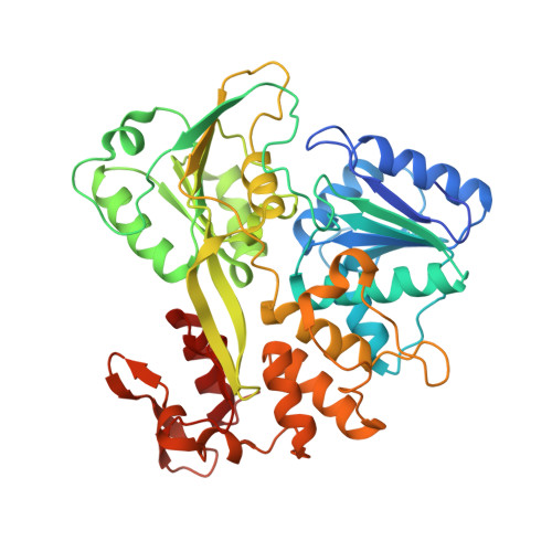
Crystallographic Snapshots of the Zika Virus NS3 Helicase Help Visualize the Reactant Water Replenishment.

Fang, J.Jing, X.Lu, G.Xu, Y.Gong, P.

(2019) ACS Infect Dis 5: 177-183

  • DOI: https://doi.org/10.1021/acsinfecdis.8b00214
  • Primary Citation of Related Structures:  
    6ADW, 6ADX, 6ADY

  • PubMed Abstract: 

    Zika virus (ZIKV), a positive-strand RNA virus belonging to the Flavivirus genus, has become an urgent public health concern since recent outbreaks worldwide. Its genome replication is facilitated by the viral NS3 protein bearing helicase function. The NS3 helicase uses energy derived from adenosine triphosphate (ATP) hydrolysis to unwind RNA duplexed regions. Structural studies of the flavivirus NS3 helicases have suggested a conserved mechanism of ATP hydrolysis. However, the process of the reactant water replenishment, a key part of the hydrolysis cycle, remains elusive. Here, we report two high-resolution crystal structures of ZIKV NS3 helicase in complex with adenosine diphosphate (ADP) and Mn 2+ , one with the reactant water already loaded as previously observed and the other with the water molecule still in a loading state. These data suggest that the reactant water replenishment can occur between the release of phosphate and the release of ADP and improves the structural basis of the NS3 ATP hydrolysis cycle.


  • Organizational Affiliation

    The Joint Center of Translational Precision Medicine, Guangzhou Institute of Pediatrics, Guangzhou Women and Children's Medical Center, No. 318 Renminzhonglu , Guangzhou , Guangdong 510623 , China.


Macromolecules
Find similar proteins by:  (by identity cutoff)  |  3D Structure
Entity ID: 1
MoleculeChains Sequence LengthOrganismDetailsImage
Serine protease NS3448Zika virusMutation(s): 0 
EC: 3.4.21.91 (PDB Primary Data), 3.6.1.15 (PDB Primary Data), 3.6.4.13 (PDB Primary Data)
UniProt
Find proteins for Q32ZE1 (Zika virus)
Explore Q32ZE1 
Go to UniProtKB:  Q32ZE1
Entity Groups  
Sequence Clusters30% Identity50% Identity70% Identity90% Identity95% Identity100% Identity
UniProt GroupQ32ZE1
Sequence Annotations
Expand
  • Reference Sequence
Experimental Data & Validation

Experimental Data

  • Method: X-RAY DIFFRACTION
  • Resolution: 1.75 Å
  • R-Value Free: 0.211 
  • R-Value Work: 0.179 
  • R-Value Observed: 0.180 
  • Space Group: P 1 21 1
Unit Cell:
Length ( Å )Angle ( ˚ )
a = 53.75α = 90
b = 68.658β = 92.5
c = 56.929γ = 90
Software Package:
Software NamePurpose
HKL-2000data scaling
PHENIXrefinement
PDB_EXTRACTdata extraction
HKL-2000data reduction
PHASERphasing

Structure Validation

View Full Validation Report


Ligand Structure Quality Assessment 


Entry History & Funding Information

Deposition Data


Funding OrganizationLocationGrant Number
Natural Science Foundation of Guangdong Province of ChinaChina2016A030310249

Revision History  (Full details and data files)

  • Version 1.0: 2019-03-13
    Type: Initial release
  • Version 1.1: 2023-11-22
    Changes: Data collection, Database references, Refinement description