6A7F

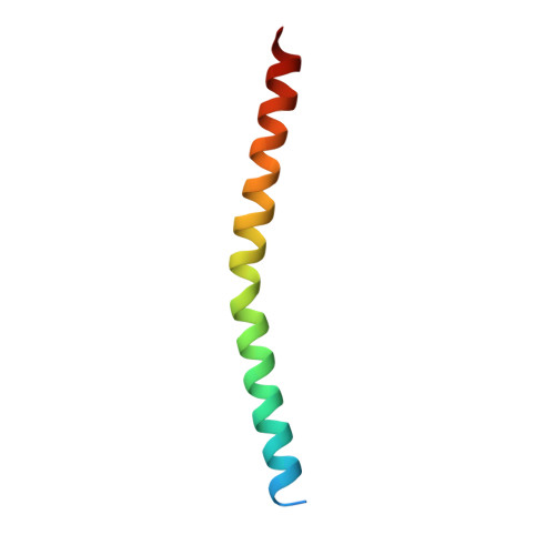
The cryo-EM structure of filamentous bacteriophage IKe major coat protein p8 shell assembly.

  • Classification: VIRAL PROTEIN
  • Organism(s): Salmonella phage IKe
  • Mutation(s): No 
  • Membrane Protein: Yes  OPM

  • Deposited: 2018-07-03 Released: 2019-02-27 
  • Deposition Author(s): Xu, J.W., Dayan, N., Goldbourt, A., Xiang, Y.
  • Funding Organization(s): Ministry of Science and Technology (China), National Basic Research Program of China (973 Program), National Natural Science Foundation of China

Experimental Data Snapshot

  • Method: ELECTRON MICROSCOPY
  • Resolution: 3.40 Å
  • Aggregation State: FILAMENT 
  • Reconstruction Method: HELICAL 

wwPDB Validation   3D Report Full Report


This is version 1.3 of the entry. See complete history


Literature

Cryo-electron microscopy structure of the filamentous bacteriophage IKe.

Xu, J.Dayan, N.Goldbourt, A.Xiang, Y.

(2019) Proc Natl Acad Sci U S A 116: 5493-5498

  • DOI: https://doi.org/10.1073/pnas.1811929116
  • Primary Citation of Related Structures:  
    6A7F

  • PubMed Abstract: 

    The filamentous bacteriophage IKe infects Escherichia coli cells bearing IncN pili. We report the cryo-electron microscopy structure of the micrometer-long IKe viral particle at a resolution of 3.4 Å. The major coat protein [protein 8 (p8)] consists of 47 residues that fold into a ∼68-Å-long helix. An atomic model of the coat protein was built. Five p8 helices in a horizontal layer form a pentamer, and symmetrically neighboring p8 layers form a right-handed helical cylinder having a rise per pentamer of 16.77 Å and a twist of 38.52°. The inner surface of the capsid cylinder is positively charged and has direct interactions with the encapsulated circular single-stranded DNA genome, which has an electron density consistent with an unusual left-handed helix structure. Similar to capsid structures of other filamentous viruses, strong capsid packing in the IKe particle is maintained by hydrophobic residues. Despite having a different length and large sequence differences from other filamentous phages, π-π interactions were found between Tyr9 of one p8 and Trp29 of a neighboring p8 in IKe that are similar to interactions observed in phage M13, suggesting that, despite sequence divergence, overall structural features are maintained.


  • Organizational Affiliation

    Center for Infectious Disease Research, School of Medicine, Tsinghua University, Beijing 100084, China.


Macromolecules
Find similar proteins by:  (by identity cutoff)  |  3D Structure
Entity ID: 1
MoleculeChains Sequence LengthOrganismDetailsImage
major coat protein p853Salmonella phage IKeMutation(s): 0 
Membrane Entity: Yes 
UniProt
Find proteins for P03620 (Salmonella phage IKe)
Explore P03620 
Go to UniProtKB:  P03620
Entity Groups  
Sequence Clusters30% Identity50% Identity70% Identity90% Identity95% Identity100% Identity
UniProt GroupP03620
Sequence Annotations
Expand
  • Reference Sequence
Experimental Data & Validation

Experimental Data

  • Method: ELECTRON MICROSCOPY
  • Resolution: 3.40 Å
  • Aggregation State: FILAMENT 
  • Reconstruction Method: HELICAL 
EM Software:
TaskSoftware PackageVersion
RECONSTRUCTIONRELION2.0
MODEL REFINEMENTPHENIX

Structure Validation

View Full Validation Report


Entry History & Funding Information

Deposition Data


Funding OrganizationLocationGrant Number
Ministry of Science and Technology (China)China2016YFA0501100
National Basic Research Program of China (973 Program)China2015CB910102
National Natural Science Foundation of ChinaChina31470721

Revision History  (Full details and data files)

  • Version 1.0: 2019-02-27
    Type: Initial release
  • Version 1.1: 2019-03-13
    Changes: Data collection, Database references
  • Version 1.2: 2019-04-03
    Changes: Data collection, Database references
  • Version 1.3: 2024-03-27
    Changes: Author supporting evidence, Data collection, Database references