5ZT8

SirB from Bacillus subtilis


Experimental Data Snapshot

  • Method: X-RAY DIFFRACTION
  • Resolution: 2.00 Å
  • R-Value Free: 0.218 
  • R-Value Work: 0.185 
  • R-Value Observed: 0.187 

wwPDB Validation   3D Report Full Report


This is version 1.3 of the entry. See complete history


Literature

Structure of sirohydrochlorin ferrochelatase SirB: the last of the structures of the class II chelatase family.

Fujishiro, T.Shimada, Y.Nakamura, R.Ooi, M.

(2019) Dalton Trans 48: 6083-6090

  • DOI: https://doi.org/10.1039/c8dt04727h
  • Primary Citation of Related Structures:  
    5ZT7, 5ZT8

  • PubMed Abstract: 

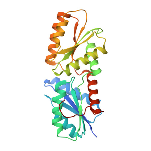
    The crystal structure of Bacillus subtilis SirB, which catalyses the insertion of Fe2+ into the substrate sirohydrochlorin (SHC) in siroheme biosynthesis, is reported herein as the last of the structures of class II chelatases. The structure of SirB with Co2+ showed that the active site of SirB is located at the N-terminal domain with metal-binding amino acid residues His10, Glu43, and His76, which was also predicted for CbiX, but is distinct from the C-terminal active sites of CbiK and HemH. The biosynthetic model reactions using SirB, Co2+ and uroporphyrin I or protoporphyrin IX as a SHC analogue revealed that SirB showed chelatase activity for uroporphyrin I, but not for protoporphyrin IX. Simulations of tetrapyrroles docking to SirB provided an insight into its tetrapyrrole substrate recognition: SHC and uroporphyrin I were suitably bound beside the Co2+ ion-binding site at the active site cavity; protoporphyrin IX was also docked to the active site but its orientation was different from those of the other two tetrapyrroles. Summarizing the present data, it was proposed that the key structural features for substrate recognition of SirB could be the hydrophobic area at the active site as well as the substituents of the tetrapyrroles.


  • Organizational Affiliation

    Department of Biochemistry and Molecular Biology, Graduate School of Science and Engineering, Saitama University, 255 Shimo-Okubo, Sakura-ku, Saitama 338-8570, Japan. tfujishiro@mail.saitama-u.ac.jp.


Macromolecules
Find similar proteins by:  (by identity cutoff)  |  3D Structure
Entity ID: 1
MoleculeChains Sequence LengthOrganismDetailsImage
Sirohydrochlorin ferrochelatase
A, B
273Bacillus subtilis subsp. subtilis str. 168Mutation(s): 0 
Gene Names: sirBylnEBSU15620
EC: 4.99.1.4
UniProt
Find proteins for O34632 (Bacillus subtilis (strain 168))
Explore O34632 
Go to UniProtKB:  O34632
Entity Groups  
Sequence Clusters30% Identity50% Identity70% Identity90% Identity95% Identity100% Identity
UniProt GroupO34632
Sequence Annotations
Expand
  • Reference Sequence
Experimental Data & Validation

Experimental Data

  • Method: X-RAY DIFFRACTION
  • Resolution: 2.00 Å
  • R-Value Free: 0.218 
  • R-Value Work: 0.185 
  • R-Value Observed: 0.187 
  • Space Group: P 1 21 1
Unit Cell:
Length ( Å )Angle ( ˚ )
a = 69.69α = 90
b = 53.37β = 105.42
c = 80.43γ = 90
Software Package:
Software NamePurpose
XSCALEdata scaling
PHENIXrefinement
PDB_EXTRACTdata extraction
XDSdata reduction
MOLREPphasing

Structure Validation

View Full Validation Report


Entry History & Funding Information

Deposition Data


Funding OrganizationLocationGrant Number
Japan Society for the Promotion of ScienceJapan17K14510

Revision History  (Full details and data files)

  • Version 1.0: 2019-02-27
    Type: Initial release
  • Version 1.1: 2019-03-06
    Changes: Data collection, Database references
  • Version 1.2: 2019-05-22
    Changes: Data collection, Database references
  • Version 1.3: 2023-11-22
    Changes: Data collection, Database references, Refinement description