


The crystal structure of inactive dimeric peptidyl-tRNA hydrolase from Acinetobacter baumannii at 1.79 A resolution
Bairagya, H.R., Sharma, P., Iqbal, N., Kaur, P., Sharma, S., Singh, T.P.To be published.
Experimental Data Snapshot
wwPDB Validation 3D Report Full Report

Entity ID: 1 | |||||
|---|---|---|---|---|---|
| Molecule | Chains | Sequence Length | Organism | Details | Image |
| Peptidyl-tRNA hydrolase | 193 | Acinetobacter baumannii ATCC 19606 = CIP 70.34 = JCM 6841 | Mutation(s): 0 Gene Names: pth, F911_03144, HMPREF0010_01329 EC: 3.1.1.29 | 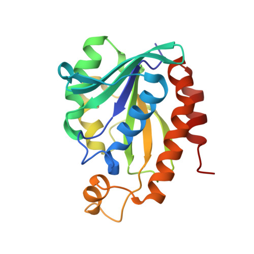 | |
UniProt | |||||
Find proteins for D0C9L6 (Acinetobacter baumannii (strain ATCC 19606 / DSM 30007 / JCM 6841 / CCUG 19606 / CIP 70.34 / NBRC 109757 / NCIMB 12457 / NCTC 12156 / 81)) Explore D0C9L6 Go to UniProtKB: D0C9L6 | |||||
Entity Groups | |||||
| Sequence Clusters | 30% Identity50% Identity70% Identity90% Identity95% Identity100% Identity | ||||
| UniProt Group | D0C9L6 | ||||
Sequence AnnotationsExpand | |||||
| |||||
| Ligands 1 Unique | |||||
|---|---|---|---|---|---|
| ID | Chains | Name / Formula / InChI Key | 2D Diagram | 3D Interactions | |
| NA Query on NA | C [auth A], D [auth B] | SODIUM ION Na FKNQFGJONOIPTF-UHFFFAOYSA-N |  | ||
| Length ( Å ) | Angle ( ˚ ) |
|---|---|
| a = 34.53 | α = 90 |
| b = 98.49 | β = 90 |
| c = 123.09 | γ = 90 |
| Software Name | Purpose |
|---|---|
| REFMAC | refinement |
| XDS | data reduction |
| SCALEPACK | data scaling |
| MOLREP | phasing |
RCSB PDB (citation) is hosted by
RCSB PDB is a member of the

