5Y7Z

Complex structure of cyclin G-associated kinase with gefitinib


Experimental Data Snapshot

  • Method: X-RAY DIFFRACTION
  • Resolution: 2.80 Å
  • R-Value Free: 0.288 
  • R-Value Work: 0.233 
  • R-Value Observed: 0.235 

wwPDB Validation   3D Report Full Report


Ligand Structure Quality Assessment 


This is version 1.2 of the entry. See complete history


Literature

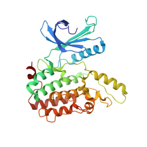
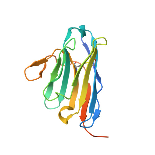
Structural Basis for the Inhibition of Cyclin G-Associated Kinase by Gefitinib.

Ohbayashi, N.Murayama, K.Kato-Murayama, M.Kukimoto-Niino, M.Uejima, T.Matsuda, T.Ohsawa, N.Yokoyoma, S.Nojima, H.Shirouzu, M.

(2018) ChemistryOpen 7: 721-727

  • DOI: https://doi.org/10.1002/open.201800177
  • Primary Citation of Related Structures:  
    5Y7Z, 5Y80

  • PubMed Abstract: 

    Gefitinib is the molecular target drug for advanced non-small-cell lung cancer. The primary target of gefitinib is the positive mutation of epidermal growth factor receptor, but it also inhibits cyclin G-associated kinase (GAK). To reveal the molecular bases of GAK and gefitinib binding, structure analyses were conducted and determined two forms of the gefitinib-bound nanobody⋅GAK kinase domain complex structures. The first form, GAK_1, has one gefitinib at the ATP binding pocket, whereas the second form, GAK_2, binds one each in the ATP binding site and a novel binding site adjacent to the activation segment C-terminal helix, a unique element of the Numb-associated kinase family. In the novel binding site, gefitinib binds in the hydrophobic groove around the activation segment, disrupting the conserved hydrogen bonds for the catalytic activity. These structures suggest possibilities for the development of selective GAK inhibitors for viral infections, such as the hepatitis C virus.


  • Organizational Affiliation

    Division of Structural and Synthetic Biology RIKEN Center for Life Science Technologies 1-7-22 Suehiro-cho, Tsurumi Yokohama 230-0045 Japan.


Macromolecules
Find similar proteins by:  (by identity cutoff)  |  3D Structure
Entity ID: 1
MoleculeChains Sequence LengthOrganismDetailsImage
Cyclin-G-associated kinase
A, B
313Homo sapiensMutation(s): 0 
Gene Names: GAK
EC: 2.7.11.1
UniProt & NIH Common Fund Data Resources
Find proteins for O14976 (Homo sapiens)
Explore O14976 
Go to UniProtKB:  O14976
PHAROS:  O14976
GTEx:  ENSG00000178950 
Entity Groups  
Sequence Clusters30% Identity50% Identity70% Identity90% Identity95% Identity100% Identity
UniProt GroupO14976
Sequence Annotations
Expand
  • Reference Sequence
Find similar proteins by:  (by identity cutoff)  |  3D Structure
Entity ID: 2
MoleculeChains Sequence LengthOrganismDetailsImage
NANOBODYC [auth D],
D [auth C]
148Lama glamaMutation(s): 0 
Entity Groups  
Sequence Clusters30% Identity50% Identity70% Identity90% Identity95% Identity100% Identity
Sequence Annotations
Expand
  • Reference Sequence
Experimental Data & Validation

Experimental Data

  • Method: X-RAY DIFFRACTION
  • Resolution: 2.80 Å
  • R-Value Free: 0.288 
  • R-Value Work: 0.233 
  • R-Value Observed: 0.235 
  • Space Group: P 21 21 21
Unit Cell:
Length ( Å )Angle ( ˚ )
a = 76.353α = 90
b = 85.513β = 90
c = 149.042γ = 90
Software Package:
Software NamePurpose
PHENIXrefinement
HKL-2000data reduction
HKL-2000data scaling
PHASERphasing

Structure Validation

View Full Validation Report


Ligand Structure Quality Assessment 


Entry History 

Deposition Data

Revision History  (Full details and data files)

  • Version 1.0: 2018-08-29
    Type: Initial release
  • Version 1.1: 2018-11-07
    Changes: Data collection, Database references
  • Version 1.2: 2023-11-22
    Changes: Data collection, Database references, Refinement description