


Structure and Catalytic Mechanism of a Novel Pyrethroid Hydrolase from Sphingobium faniae JZ-2
Xu, D.Q., Gao, Y.Y., Ran, T.T., Zeng, L.P., He, J., Wang, W.W.To be published.
Experimental Data Snapshot
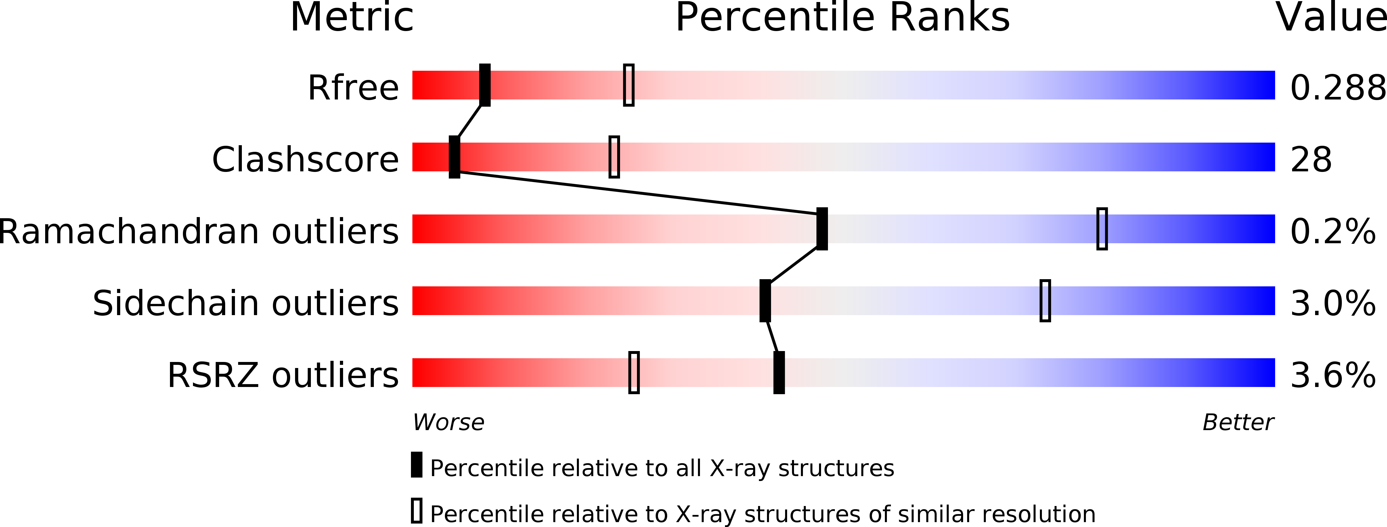
wwPDB Validation 3D Report Full Report
Entity ID: 1 | |||||
|---|---|---|---|---|---|
| Molecule | Chains | Sequence Length | Organism | Details | Image |
| Pyrethroid hydrolase | 288 | Sphingobium faniae | Mutation(s): 1 Gene Names: pytH | 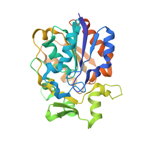 | |
UniProt | |||||
Find proteins for D0VUS3 (Sphingobium faniae) Explore D0VUS3 Go to UniProtKB: D0VUS3 | |||||
Entity Groups | |||||
| Sequence Clusters | 30% Identity50% Identity70% Identity90% Identity95% Identity100% Identity | ||||
| UniProt Group | D0VUS3 | ||||
Sequence AnnotationsExpand | |||||
| |||||
| Ligands 1 Unique | |||||
|---|---|---|---|---|---|
| ID | Chains | Name / Formula / InChI Key | 2D Diagram | 3D Interactions | |
| SO4 Query on SO4 | G [auth A] H [auth B] I [auth C] J [auth D] K [auth E] | SULFATE ION O4 S QAOWNCQODCNURD-UHFFFAOYSA-L |  | ||
| Length ( Å ) | Angle ( ˚ ) |
|---|---|
| a = 168.2 | α = 90 |
| b = 168.2 | β = 90 |
| c = 123.626 | γ = 90 |
| Software Name | Purpose |
|---|---|
| PHENIX | refinement |
| XDS | data reduction |
| SCALA | data scaling |
| PHASER | phasing |
| Funding Organization | Location | Grant Number |
|---|---|---|
| State's Key Project of Research and Development Plan | China | 2016YFD0801102 |
| Natural Science Foundation of China | China | 31400055 |
RCSB PDB (citation) is hosted by
RCSB PDB is a member of the

