5Y42

Native-crystal structure of three chain non-toxic type II ribosome inactivating protein purified from the seeds of Trichosanthes anguina


Experimental Data Snapshot

  • Method: X-RAY DIFFRACTION
  • Resolution: 2.90 Å
  • R-Value Free: 0.255 
  • R-Value Work: 0.202 
  • R-Value Observed: 0.205 

wwPDB Validation   3D Report Full Report


Ligand Structure Quality Assessment 


This is version 1.3 of the entry. See complete history


Literature

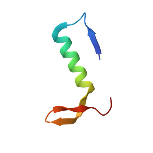
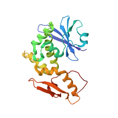
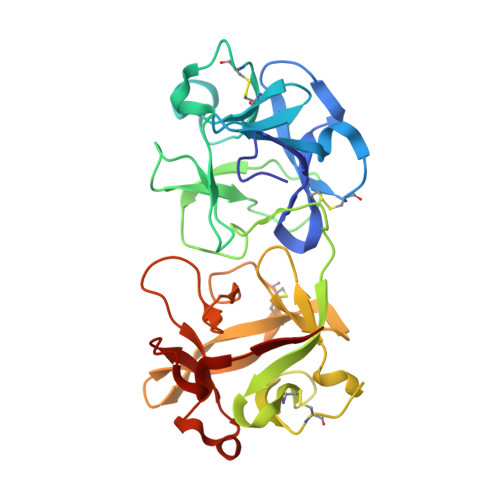
Ligand binding and retention in snake gourd seed lectin (SGSL). A crystallographic, thermodynamic and molecular dynamics study.

Chandran, T.Sivaji, N.Surolia, A.Vijayan, M.

(2018) Glycobiology 28: 968-977

  • DOI: https://doi.org/10.1093/glycob/cwy072
  • Primary Citation of Related Structures:  
    5Y42, 5Y97

  • PubMed Abstract: 

    Snake gourd seed lectin (SGSL) is a non-toxic homolog of type II ribosome-inactivating proteins (RIPs) which contain a catalytic domain and a lectin domain. Isothermal titration calorimetry (ITC) measurements of the interactions of the protein with LacNAc, Lac, Gal, Me-α-Gal were carried out and the crystal structures of the native protein and its complex with Lac were determined. The crystal structure of the Me-α-Gal complex has already been determined. While the crystal structure showed the presence of two-sugar-binding sites, one on each of the two domains of the lectin chain, ITC measurements indicated the presence of only one binding site. In order to resolve this anomaly, molecular dynamics (MD) simulations were carried out on the native protein and on its complexes with Me-α-Gal and Lac. Simulations were also performed on the protein after reducing the inter-chain disulfide bridge between the two chains. The crystal structures and the simulations confirmed the robustness of the protein structure, irrespective of the presence or absence of the disulfide bridge. The simulations indicated that although two sites can bind sugar, only the ligand at one site is retained in a dynamic situation. The studies thus bring out the subtle relationship between binding and retention of the ligand.


  • Organizational Affiliation

    Molecular Biophysics Unit, Indian Institute of Science, Bangalore, India.


Macromolecules
Find similar proteins by:  (by identity cutoff)  |  3D Structure
Entity ID: 1
MoleculeChains Sequence LengthOrganismDetailsImage
Lectin
A, D
41Trichosanthes anguinaMutation(s): 0 
UniProt
Find proteins for U3KRF8 (Trichosanthes anguina)
Explore U3KRF8 
Go to UniProtKB:  U3KRF8
Entity Groups  
Sequence Clusters30% Identity50% Identity70% Identity90% Identity95% Identity100% Identity
UniProt GroupU3KRF8
Sequence Annotations
Expand
  • Reference Sequence
Find similar proteins by:  (by identity cutoff)  |  3D Structure
Entity ID: 2
MoleculeChains Sequence LengthOrganismDetailsImage
Seed lectin
B, E
206Trichosanthes anguinaMutation(s): 0 
UniProt
Find proteins for U3KRF8 (Trichosanthes anguina)
Explore U3KRF8 
Go to UniProtKB:  U3KRF8
Entity Groups  
Sequence Clusters30% Identity50% Identity70% Identity90% Identity95% Identity100% Identity
UniProt GroupU3KRF8
Sequence Annotations
Expand
  • Reference Sequence
Find similar proteins by:  (by identity cutoff)  |  3D Structure
Entity ID: 3
MoleculeChains Sequence LengthOrganismDetailsImage
Seed lectin
C, F
264Trichosanthes anguinaMutation(s): 0 
UniProt
Find proteins for U3KRF8 (Trichosanthes anguina)
Explore U3KRF8 
Go to UniProtKB:  U3KRF8
Entity Groups  
Sequence Clusters30% Identity50% Identity70% Identity90% Identity95% Identity100% Identity
UniProt GroupU3KRF8
Sequence Annotations
Expand
  • Reference Sequence
Small Molecules
Ligands 1 Unique
IDChains Name / Formula / InChI Key2D Diagram3D Interactions
NAG
Query on NAG

Download Ideal Coordinates CCD File 
G [auth C]2-acetamido-2-deoxy-beta-D-glucopyranose
C8 H15 N O6
OVRNDRQMDRJTHS-FMDGEEDCSA-N
Experimental Data & Validation

Experimental Data

  • Method: X-RAY DIFFRACTION
  • Resolution: 2.90 Å
  • R-Value Free: 0.255 
  • R-Value Work: 0.202 
  • R-Value Observed: 0.205 
  • Space Group: P 64
Unit Cell:
Length ( Å )Angle ( ˚ )
a = 171.226α = 90
b = 171.226β = 90
c = 75.85γ = 120
Software Package:
Software NamePurpose
REFMACrefinement
iMOSFLMdata reduction
SCALAdata scaling
PHASERphasing

Structure Validation

View Full Validation Report


Ligand Structure Quality Assessment 


Entry History & Funding Information

Deposition Data


Funding OrganizationLocationGrant Number
SERB, CSIRIndia--

Revision History  (Full details and data files)

  • Version 1.0: 2018-08-08
    Type: Initial release
  • Version 1.1: 2019-02-20
    Changes: Data collection, Database references
  • Version 1.2: 2020-07-29
    Type: Remediation
    Reason: Carbohydrate remediation
    Changes: Data collection, Derived calculations, Structure summary
  • Version 1.3: 2023-11-22
    Changes: Data collection, Database references, Refinement description, Structure summary