5Y1I

The crystal structure of GfsF


Experimental Data Snapshot

  • Method: X-RAY DIFFRACTION
  • Resolution: 2.00 Å
  • R-Value Free: 0.236 
  • R-Value Work: 0.194 
  • R-Value Observed: 0.196 

wwPDB Validation   3D Report Full Report


Ligand Structure Quality Assessment 


This is version 1.2 of the entry. See complete history


Literature

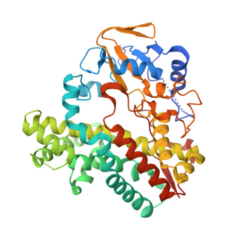
Substrate Recognition by a Dual-Function P450 Monooxygenase GfsF Involved in FD-891 Biosynthesis

Miyanaga, A.Takayanagi, R.Furuya, T.Kawamata, A.Itagaki, T.Iwabuchi, Y.Kanoh, N.Kudo, F.Eguchi, T.

(2017) Chembiochem 18: 2179-2187

  • DOI: https://doi.org/10.1002/cbic.201700429
  • Primary Citation of Related Structures:  
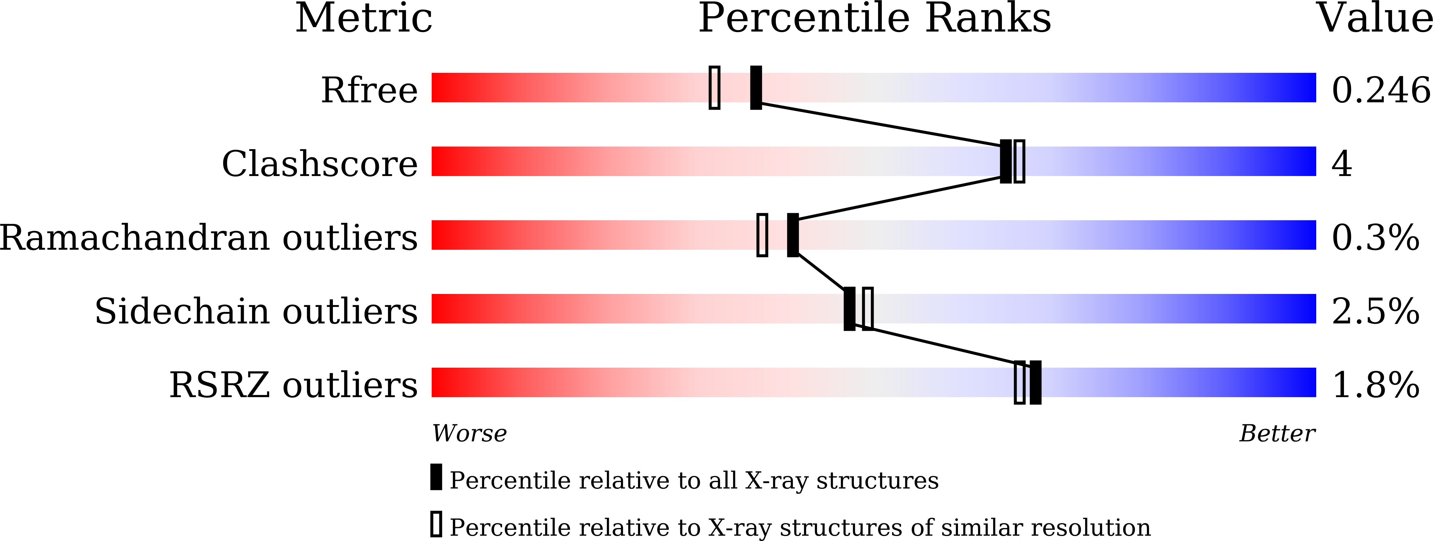
    5Y1I

  • PubMed Abstract: 

    GfsF is a multifunctional P450 monooxygenase that catalyzes epoxidation and subsequent hydroxylation in the biosynthesis of macrolide polyketide FD-891. Here, we describe the biochemical and structural analysis of GfsF. To obtain the structural basis of a dual-function reaction, we determined the crystal structure of ligand-free GfsF, which revealed GfsF to have a predominantly hydrophobic substrate binding pocket. The docking models, in conjunction with the results of the enzymatic assay with substrate analogues and site-directed mutagenesis suggested two distinct substrate binding modes for epoxidation and hydroxylation reactions, which explained how GfsF regulates the order of two oxidative reactions. These findings provide new insights into the reaction mechanism of multifunctional P450 monooxygenases.


  • Organizational Affiliation

    Department of Chemistry, Tokyo Institute of Technology, 2-12-1 O-okayama, Meguro-ku, Tokyo, 152-8551, Japan.


Macromolecules
Find similar proteins by:  (by identity cutoff)  |  3D Structure
Entity ID: 1
MoleculeChains Sequence LengthOrganismDetailsImage
Cytochrome P450
A, B
403Streptomyces graminofaciensMutation(s): 0 
Gene Names: gfsF
UniProt
Find proteins for E0D207 (Streptomyces graminofaciens)
Explore E0D207 
Go to UniProtKB:  E0D207
Entity Groups  
Sequence Clusters30% Identity50% Identity70% Identity90% Identity95% Identity100% Identity
UniProt GroupE0D207
Sequence Annotations
Expand
  • Reference Sequence
Experimental Data & Validation

Experimental Data

  • Method: X-RAY DIFFRACTION
  • Resolution: 2.00 Å
  • R-Value Free: 0.236 
  • R-Value Work: 0.194 
  • R-Value Observed: 0.196 
  • Space Group: P 21 21 21
Unit Cell:
Length ( Å )Angle ( ˚ )
a = 67.08α = 90
b = 102.87β = 90
c = 114.43γ = 90
Software Package:
Software NamePurpose
REFMACrefinement
iMOSFLMdata reduction
SCALAdata scaling
MOLREPphasing

Structure Validation

View Full Validation Report


Ligand Structure Quality Assessment 


Entry History & Funding Information

Deposition Data


Funding OrganizationLocationGrant Number
Agricultural Chemical Research FoundationJapan40-5

Revision History  (Full details and data files)

  • Version 1.0: 2017-09-13
    Type: Initial release
  • Version 1.1: 2017-11-15
    Changes: Database references
  • Version 1.2: 2023-11-22
    Changes: Data collection, Database references, Derived calculations, Refinement description