5XNX

Crystallographic structure of the enzymatically active N-terminal domain of the Rel protein from Mycobacterium tuberculosis


Experimental Data Snapshot

  • Method: X-RAY DIFFRACTION
  • Resolution: 3.70 Å
  • R-Value Free: 0.368 
  • R-Value Work: 0.350 
  • R-Value Observed: 0.351 

wwPDB Validation   3D Report Full Report


This is version 1.2 of the entry. See complete history


Literature

Crystallographic and solution structure of the N-terminal domain of the Rel protein from Mycobacterium tuberculosis

Singal, B.Balakrishna, A.M.Nartey, W.Manimekalai, M.S.S.Jeyakanthan, J.Gruber, G.

(2017) FEBS Lett 591: 2323-2337

  • DOI: https://doi.org/10.1002/1873-3468.12739
  • Primary Citation of Related Structures:  
    5XNX

  • PubMed Abstract: 

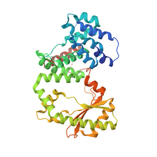
    Modulation of intracellular guanosine 3',5'-bispyrophosphate ((p)ppGpp) level, the effector of the stringent response, is crucial for survival as well as optimal growth of prokaryotes and, thus, for bacterial pathogenesis and dormancy. In Mycobacterium tuberculosis (Mtb), (p)ppGpp synthesis and degradation are carried out by the bifunctional enzyme MtRel, which consists of 738 residues, including an N-terminal hydrolase- and synthetase-domain (N-terminal domain or NTD) and a C-terminus with a ribosome-binding site. Here, we present the first crystallographic structure of the enzymatically active MtRel NTD determined at 3.7 Å resolution. The structure provides insights into the residues of MtRel NTD responsible for nucleotide binding. Small-angle X-ray scattering experiments were performed to investigate the dimeric state of the MtRel NTD and possible substrate-dependent structural alterations.


  • Organizational Affiliation

    School of Biological Sciences, Nanyang Technological University, Singapore, Singapore.


Macromolecules
Find similar proteins by:  (by identity cutoff)  |  3D Structure
Entity ID: 1
MoleculeChains Sequence LengthOrganismDetailsImage
Bifunctional (p)ppGpp synthase/hydrolase RelA
A, B, C, D
394Mycobacterium tuberculosis H37RvMutation(s): 0 
Gene Names: relARv2583cMTCY227.18
EC: 2.7.6.5 (PDB Primary Data), 3.1.7.2 (PDB Primary Data)
UniProt
Find proteins for P9WHG9 (Mycobacterium tuberculosis (strain ATCC 25618 / H37Rv))
Explore P9WHG9 
Go to UniProtKB:  P9WHG9
Entity Groups  
Sequence Clusters30% Identity50% Identity70% Identity90% Identity95% Identity100% Identity
UniProt GroupP9WHG9
Sequence Annotations
Expand
  • Reference Sequence
Experimental Data & Validation

Experimental Data

  • Method: X-RAY DIFFRACTION
  • Resolution: 3.70 Å
  • R-Value Free: 0.368 
  • R-Value Work: 0.350 
  • R-Value Observed: 0.351 
  • Space Group: P 32
Unit Cell:
Length ( Å )Angle ( ˚ )
a = 161.71α = 90
b = 161.71β = 90
c = 75.56γ = 120
Software Package:
Software NamePurpose
REFMACrefinement
HKL-2000data reduction
HKL-2000data scaling
PHASERphasing

Structure Validation

View Full Validation Report


Entry History 

Deposition Data

Revision History  (Full details and data files)

  • Version 1.0: 2017-07-19
    Type: Initial release
  • Version 1.1: 2017-08-30
    Changes: Database references
  • Version 1.2: 2023-11-22
    Changes: Data collection, Database references, Refinement description