5XJ5

Crystal structure of PlsY (YgiH), an integral membrane glycerol 3-phosphate acyltransferase - the monoacylglycerol form

  • Classification: TRANSFERASE
  • Organism(s): Aquifex aeolicus VF5
  • Expression System: Escherichia coli BL21(DE3)
  • Mutation(s): No 
  • Membrane Protein: Yes  OPMPDBTM

  • Deposited: 2017-04-30 Released: 2017-12-06 
  • Deposition Author(s): Li, Z., Li, D.
  • Funding Organization(s): National Natural Science Foundation of China, Chinese Academy of Sciences (CAS)-Shanghai Science Research Center, Key Program of CAS Frontier Science, 1000 Young Talent Program, Shanghai Pujiang Talent Program

Experimental Data Snapshot

  • Method: X-RAY DIFFRACTION
  • Resolution: 1.48 Å
  • R-Value Free: 0.213 
  • R-Value Work: 0.188 
  • R-Value Observed: 0.189 

wwPDB Validation   3D Report Full Report


Ligand Structure Quality Assessment 


This is version 1.0 of the entry. See complete history


Literature

Structural insights into the committed step of bacterial phospholipid biosynthesis.

Li, Z.Tang, Y.Wu, Y.Zhao, S.Bao, J.Luo, Y.Li, D.

(2017) Nat Commun 8: 1691-1691

  • DOI: https://doi.org/10.1038/s41467-017-01821-9
  • Primary Citation of Related Structures:  
    5XJ5, 5XJ6, 5XJ7, 5XJ8, 5XJ9

  • PubMed Abstract: 

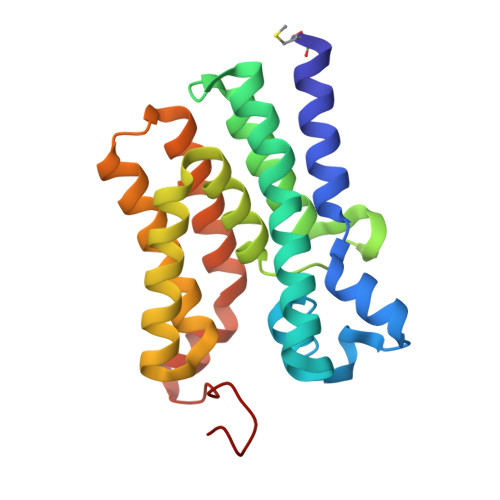
    The membrane-integral glycerol 3-phosphate (G3P) acyltransferase PlsY catalyses the committed and essential step in bacterial phospholipid biosynthesis by acylation of G3P, forming lysophosphatidic acid. It contains no known acyltransferase motifs, lacks eukaryotic homologs, and uses the unusual acyl-phosphate as acyl donor, as opposed to acyl-CoA or acyl-carrier protein for other acyltransferases. Previous studies have identified several PlsY inhibitors as potential antimicrobials. Here we determine the crystal structure of PlsY at 1.48 Å resolution, revealing a seven-transmembrane helix fold. Four additional substrate- and product-bound structures uncover the atomic details of its relatively inflexible active site. Structure and mutagenesis suggest a different acylation mechanism of 'substrate-assisted catalysis' that, unlike other acyltransferases, does not require a proteinaceous catalytic base to complete. The structure data and a high-throughput enzymatic assay developed in this work should prove useful for virtual and experimental screening of inhibitors against this vital bacterial enzyme.


  • Organizational Affiliation

    State Key Laboratory of Molecular Biology, National Center for Protein Science Shanghai, Shanghai Science Research Center, CAS Center for Excellence in Molecular Cell Science, Shanghai Institute of Biochemistry and Cell Biology, Chinese Academy of Sciences, 333 Haike Road, Shanghai, 201210, China.


Macromolecules
Find similar proteins by:  (by identity cutoff)  |  3D Structure
Entity ID: 1
MoleculeChains Sequence LengthOrganismDetailsImage
Glycerol-3-phosphate acyltransferase201Aquifex aeolicus VF5Mutation(s): 0 
Gene Names: plsYaq_676
EC: 2.3.1
Membrane Entity: Yes 
UniProt
Find proteins for O66905 (Aquifex aeolicus (strain VF5))
Explore O66905 
Go to UniProtKB:  O66905
Entity Groups  
Sequence Clusters30% Identity50% Identity70% Identity90% Identity95% Identity100% Identity
UniProt GroupO66905
Sequence Annotations
Expand
  • Reference Sequence
Small Molecules
Ligands 4 Unique
IDChains Name / Formula / InChI Key2D Diagram3D Interactions
78M
Query on 78M

Download Ideal Coordinates CCD File 
I [auth A]
J [auth A]
K [auth A]
L [auth A]
M [auth A]
I [auth A],
J [auth A],
K [auth A],
L [auth A],
M [auth A],
N [auth A],
O [auth A],
P [auth A],
Q [auth A],
R [auth A],
S [auth A],
T [auth A],
U [auth A],
V [auth A],
W [auth A]
(2S)-2,3-DIHYDROXYPROPYL(7Z)-PENTADEC-7-ENOATE
C18 H34 O4
BJMLBVHMHXYQFS-JJEJIETFSA-N
PGE
Query on PGE

Download Ideal Coordinates CCD File 
X [auth A]TRIETHYLENE GLYCOL
C6 H14 O4
ZIBGPFATKBEMQZ-UHFFFAOYSA-N
SO4
Query on SO4

Download Ideal Coordinates CCD File 
B [auth A]
C [auth A]
D [auth A]
E [auth A]
F [auth A]
B [auth A],
C [auth A],
D [auth A],
E [auth A],
F [auth A],
G [auth A],
H [auth A]
SULFATE ION
O4 S
QAOWNCQODCNURD-UHFFFAOYSA-L
GLY
Query on GLY

Download Ideal Coordinates CCD File 
Y [auth A]GLYCINE
C2 H5 N O2
DHMQDGOQFOQNFH-UHFFFAOYSA-N
Modified Residues  1 Unique
IDChains TypeFormula2D DiagramParent
FME
Query on FME
A
L-PEPTIDE LINKINGC6 H11 N O3 SMET
Experimental Data & Validation

Experimental Data

  • Method: X-RAY DIFFRACTION
  • Resolution: 1.48 Å
  • R-Value Free: 0.213 
  • R-Value Work: 0.188 
  • R-Value Observed: 0.189 
  • Space Group: I 2 2 2
Unit Cell:
Length ( Å )Angle ( ˚ )
a = 57.675α = 90
b = 83.437β = 90
c = 107.588γ = 90
Software Package:
Software NamePurpose
PHENIXrefinement
XDSdata reduction
XDSdata scaling
PHENIXphasing

Structure Validation

View Full Validation Report


Ligand Structure Quality Assessment 


Entry History & Funding Information

Deposition Data

  • Released Date: 2017-12-06 
  • Deposition Author(s): Li, Z., Li, D.

Funding OrganizationLocationGrant Number
National Natural Science Foundation of ChinaChina31570748
National Natural Science Foundation of ChinaChinaU1632127
Chinese Academy of Sciences (CAS)-Shanghai Science Research CenterChinaCAS-SSRC-YJ-2015-02
Key Program of CAS Frontier ScienceChinaQYZDB-SSW-SMC037
1000 Young Talent ProgramChina2015
Shanghai Pujiang Talent ProgramChina15PJ1409400

Revision History  (Full details and data files)

  • Version 1.0: 2017-12-06
    Type: Initial release