5X2L

Crystal Structure of Human Serine Racemase


Experimental Data Snapshot

  • Method: X-RAY DIFFRACTION
  • Resolution: 1.81 Å
  • R-Value Free: 0.258 
  • R-Value Work: 0.220 
  • R-Value Observed: 0.222 

wwPDB Validation   3D Report Full Report


Ligand Structure Quality Assessment 


This is version 1.1 of the entry. See complete history


Literature

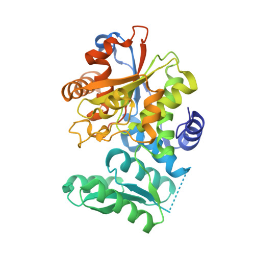
Design, synthesis, and evaluation of novel inhibitors for wild-type human serine racemase.

Takahara, S.Nakagawa, K.Uchiyama, T.Yoshida, T.Matsumoto, K.Kawasumi, Y.Mizuguchi, M.Obita, T.Watanabe, Y.Hayakawa, D.Gouda, H.Mori, H.Toyooka, N.

(2017) Bioorg Med Chem Lett 

  • DOI: https://doi.org/10.1016/j.bmcl.2017.12.021
  • Primary Citation of Related Structures:  
    5X2L

  • PubMed Abstract: 

    Most of the endogenous free d-serine (about 90%) in the brain is produced by serine racemase (SR). d-Serine in the brain is involved in neurodegenerative disorders and epileptic states as an endogenous co-agonist of the NMDA-type glutamate receptor. Thus, SR inhibitors are expected to be novel therapeutic candidates for the treatment of these disorders. In this study, we solved the crystal structure of wild-type SR, and tried to identify a new inhibitor of SR by in silico screening using the structural information. As a result, we identified two hit compounds by their in vitro evaluations using wild-type SR. Based on the structure of the more potent hit compound 1, we synthesized 15 derivatives and evaluated their inhibitory activities against wild-type SR. Among them, the compound 9C showed relatively high inhibitory potency for wild-type SR. Compound 9C was a more potent inhibitor than compound 24, which was synthesized by our group based upon the structural information of the mutant-type SR.


  • Organizational Affiliation

    Graduate School of Innovative Life Science, University of Toyama, Japan.


Macromolecules
Find similar proteins by:  (by identity cutoff)  |  3D Structure
Entity ID: 1
MoleculeChains Sequence LengthOrganismDetailsImage
Serine racemase
A, B
348Homo sapiensMutation(s): 0 
Gene Names: SRR
EC: 5.1.1.18 (PDB Primary Data), 4.3.1.18 (PDB Primary Data), 4.3.1.17 (PDB Primary Data)
UniProt & NIH Common Fund Data Resources
Find proteins for Q9GZT4 (Homo sapiens)
Explore Q9GZT4 
Go to UniProtKB:  Q9GZT4
PHAROS:  Q9GZT4
GTEx:  ENSG00000167720 
Entity Groups  
Sequence Clusters30% Identity50% Identity70% Identity90% Identity95% Identity100% Identity
UniProt GroupQ9GZT4
Sequence Annotations
Expand
  • Reference Sequence
Experimental Data & Validation

Experimental Data

  • Method: X-RAY DIFFRACTION
  • Resolution: 1.81 Å
  • R-Value Free: 0.258 
  • R-Value Work: 0.220 
  • R-Value Observed: 0.222 
  • Space Group: P 21 21 2
Unit Cell:
Length ( Å )Angle ( ˚ )
a = 80.12α = 90
b = 112.59β = 90
c = 88γ = 90
Software Package:
Software NamePurpose
PHENIXrefinement
iMOSFLMdata reduction
SCALAdata scaling
PHASERphasing

Structure Validation

View Full Validation Report


Ligand Structure Quality Assessment 


Entry History 

Deposition Data

Revision History  (Full details and data files)

  • Version 1.0: 2018-01-31
    Type: Initial release
  • Version 1.1: 2021-02-24
    Changes: Derived calculations