


Crystal structure of Phosphate-binding protein PstS protein from Burkholderia pseudomallei
Abendroth, J., Dranow, D.M., Lorimer, D.D., Edwards, T.E.To be published.
Experimental Data Snapshot
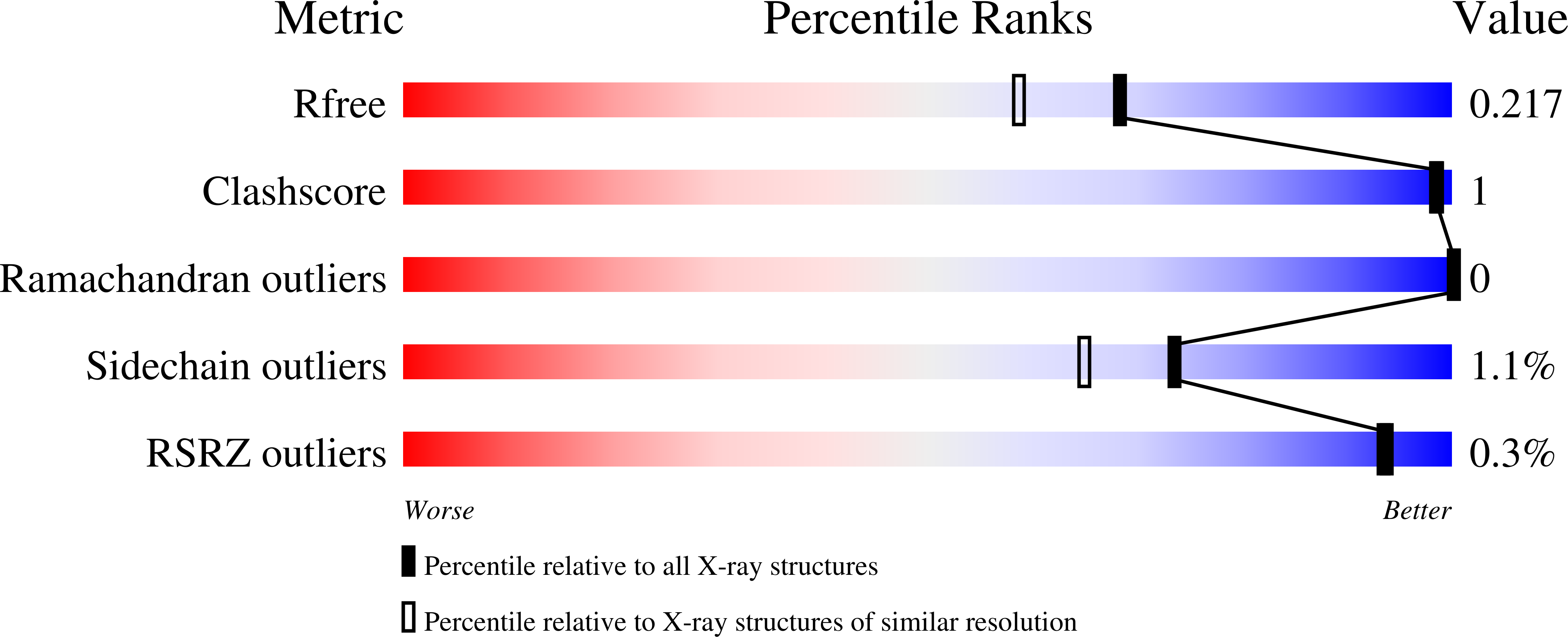
wwPDB Validation 3D Report Full Report
Entity ID: 1 | |||||
|---|---|---|---|---|---|
| Molecule | Chains | Sequence Length | Organism | Details | Image |
| Phosphate-binding protein PstS | 363 | Burkholderia pseudomallei K96243 | Mutation(s): 0 Gene Names: pstS, BPSL1359 | 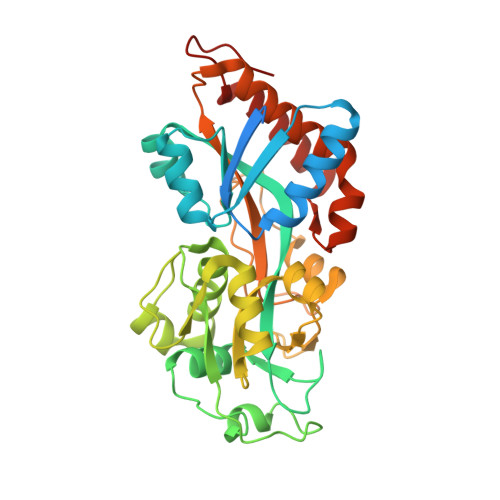 | |
UniProt | |||||
Find proteins for Q63V82 (Burkholderia pseudomallei (strain K96243)) Explore Q63V82 Go to UniProtKB: Q63V82 | |||||
Entity Groups | |||||
| Sequence Clusters | 30% Identity50% Identity70% Identity90% Identity95% Identity100% Identity | ||||
| UniProt Group | Q63V82 | ||||
Sequence AnnotationsExpand | |||||
| |||||
| Ligands 2 Unique | |||||
|---|---|---|---|---|---|
| ID | Chains | Name / Formula / InChI Key | 2D Diagram | 3D Interactions | |
| PO4 Query on PO4 | C [auth A], E [auth B] | PHOSPHATE ION O4 P NBIIXXVUZAFLBC-UHFFFAOYSA-K |  | ||
| K Query on K | D [auth A], F [auth B] | POTASSIUM ION K NPYPAHLBTDXSSS-UHFFFAOYSA-N |  | ||
| Length ( Å ) | Angle ( ˚ ) |
|---|---|
| a = 116.64 | α = 90 |
| b = 59.84 | β = 120.97 |
| c = 101.09 | γ = 90 |
| Software Name | Purpose |
|---|---|
| XDS | data reduction |
| XSCALE | data scaling |
| PHASER | phasing |
| Coot | model building |
| PHENIX | refinement |
| PDB_EXTRACT | data extraction |
RCSB PDB (citation) is hosted by
RCSB PDB is a member of the

