5WCU

Crystal structure of 167 bp nucleosome bound to the globular domain of linker histone H5


Experimental Data Snapshot

  • Method: X-RAY DIFFRACTION
  • Resolution: 5.53 Å
  • R-Value Free: 0.238 
  • R-Value Work: 0.189 
  • R-Value Observed: 0.195 

wwPDB Validation   3D Report Full Report


This is version 1.1 of the entry. See complete history


Literature

Revisit of Reconstituted 30-nm Nucleosome Arrays Reveals an Ensemble of Dynamic Structures.

Zhou, B.R.Jiang, J.Ghirlando, R.Norouzi, D.Sathish Yadav, K.N.Feng, H.Wang, R.Zhang, P.Zhurkin, V.Bai, Y.

(2018) J Mol Biol 430: 3093-3110

  • DOI: https://doi.org/10.1016/j.jmb.2018.06.020
  • Primary Citation of Related Structures:  
    5WCU

  • PubMed Abstract: 

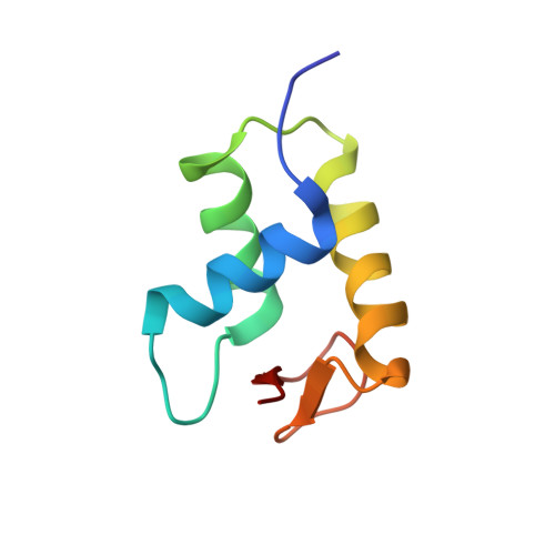
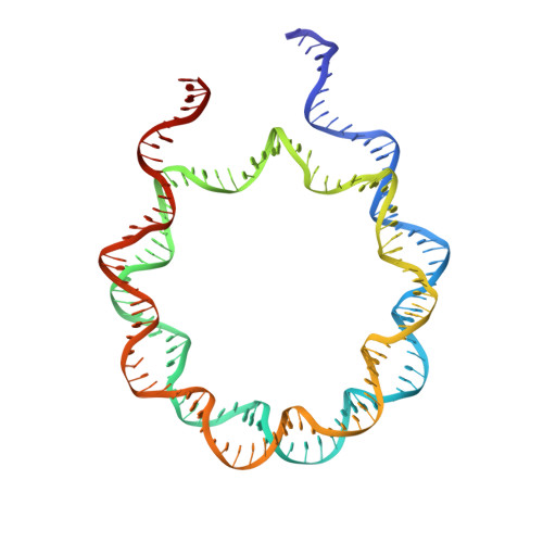
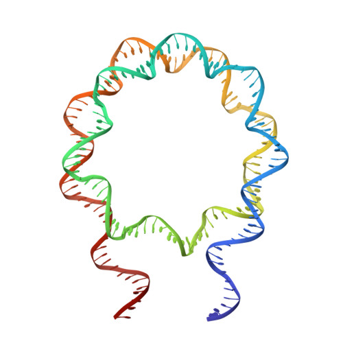
    It has long been suggested that chromatin may form a fiber with a diameter of ~30 nm that suppresses transcription. Despite nearly four decades of study, the structural nature of the 30-nm chromatin fiber and conclusive evidence of its existence in vivo remain elusive. The key support for the existence of specific 30-nm chromatin fiber structures is based on the determination of the structures of reconstituted nucleosome arrays using X-ray crystallography and single-particle cryo-electron microscopy coupled with glutaraldehyde chemical cross-linking. Here we report the characterization of these nucleosome arrays in solution using analytical ultracentrifugation, NMR, and small-angle X-ray scattering. We found that the physical properties of these nucleosome arrays in solution are not consistent with formation of just a few discrete structures of nucleosome arrays. In addition, we obtained a crystal of the nucleosome in complex with the globular domain of linker histone H5 that shows a new form of nucleosome packing and suggests a plausible alternative compact conformation for nucleosome arrays. Taken together, our results challenge the key evidence for the existence of a limited number of structures of reconstituted nucleosome arrays in solution by revealing that the reconstituted nucleosome arrays are actually best described as an ensemble of various conformations with a zigzagged arrangement of nucleosomes. Our finding has implications for understanding the structure and function of chromatin in vivo.


  • Organizational Affiliation

    Laboratory of Biochemistry and Molecular Biology, National Cancer Institute, National Institutes of Health, Bethesda, MD 20892, USA.


Macromolecules

Find similar proteins by:  (by identity cutoff)  |  3D Structure
Entity ID: 1
MoleculeChains Sequence LengthOrganismDetailsImage
Histone H3
A, E, K, O
98Drosophila melanogasterMutation(s): 0 
Gene Names: 
UniProt
Find proteins for P02299 (Drosophila melanogaster)
Explore P02299 
Go to UniProtKB:  P02299
Entity Groups  
Sequence Clusters30% Identity50% Identity70% Identity90% Identity95% Identity100% Identity
UniProt GroupP02299
Sequence Annotations
Expand
  • Reference Sequence
Find similar proteins by:  (by identity cutoff)  |  3D Structure
Entity ID: 2
MoleculeChains Sequence LengthOrganismDetailsImage
Histone H4
B, F, L, P
82Drosophila melanogasterMutation(s): 0 
Gene Names: 
UniProt
Find proteins for P84040 (Drosophila melanogaster)
Explore P84040 
Go to UniProtKB:  P84040
Entity Groups  
Sequence Clusters30% Identity50% Identity70% Identity90% Identity95% Identity100% Identity
UniProt GroupP84040
Sequence Annotations
Expand
  • Reference Sequence
Find similar proteins by:  (by identity cutoff)  |  3D Structure
Entity ID: 3
MoleculeChains Sequence LengthOrganismDetailsImage
Histone H2A
C, G, M, Q
104Drosophila melanogasterMutation(s): 0 
Gene Names: 
UniProt
Find proteins for P84051 (Drosophila melanogaster)
Explore P84051 
Go to UniProtKB:  P84051
Entity Groups  
Sequence Clusters30% Identity50% Identity70% Identity90% Identity95% Identity100% Identity
UniProt GroupP84051
Sequence Annotations
Expand
  • Reference Sequence
Find similar proteins by:  (by identity cutoff)  |  3D Structure
Entity ID: 4
MoleculeChains Sequence LengthOrganismDetailsImage
Histone H2B
D, H, N, R
94Drosophila melanogasterMutation(s): 0 
Gene Names: 
UniProt
Find proteins for P02283 (Drosophila melanogaster)
Explore P02283 
Go to UniProtKB:  P02283
Entity Groups  
Sequence Clusters30% Identity50% Identity70% Identity90% Identity95% Identity100% Identity
UniProt GroupP02283
Sequence Annotations
Expand
  • Reference Sequence
Find similar proteins by:  (by identity cutoff)  |  3D Structure
Entity ID: 7
MoleculeChains Sequence LengthOrganismDetailsImage
Histone H5
U, V
76Gallus gallusMutation(s): 0 
UniProt
Find proteins for P02259 (Gallus gallus)
Explore P02259 
Go to UniProtKB:  P02259
Entity Groups  
Sequence Clusters30% Identity50% Identity70% Identity90% Identity95% Identity100% Identity
UniProt GroupP02259
Sequence Annotations
Expand
  • Reference Sequence
Find similar nucleic acids by:  (by identity cutoff)  |  3D Structure
Entity ID: 5
MoleculeChains LengthOrganismImage
DNA (167-MER)
I, S
167synthetic construct
Sequence Annotations
Expand
  • Reference Sequence
Find similar nucleic acids by:  (by identity cutoff)  |  3D Structure
Entity ID: 6
MoleculeChains LengthOrganismImage
DNA (167-MER)
J, T
167synthetic construct
Sequence Annotations
Expand
  • Reference Sequence
Experimental Data & Validation

Experimental Data

  • Method: X-RAY DIFFRACTION
  • Resolution: 5.53 Å
  • R-Value Free: 0.238 
  • R-Value Work: 0.189 
  • R-Value Observed: 0.195 
  • Space Group: P 1
Unit Cell:
Length ( Å )Angle ( ˚ )
a = 65.926α = 100.79
b = 108.543β = 90.08
c = 180.77γ = 89.94
Software Package:
Software NamePurpose
PHENIXrefinement
XDSdata reduction
XDSdata scaling
PHASERphasing

Structure Validation

View Full Validation Report


Entry History 

Deposition Data

Revision History  (Full details and data files)

  • Version 1.0: 2018-10-31
    Type: Initial release
  • Version 1.1: 2023-10-04
    Changes: Data collection, Database references, Refinement description