


Crystal structure of DNA polymerase III subunit beta from Rickettsia conorii
Bowatte, K., Conrady, D.G., Abendroth, J., Lorimer, D.D., Edwards, T.E.To be published.
Experimental Data Snapshot
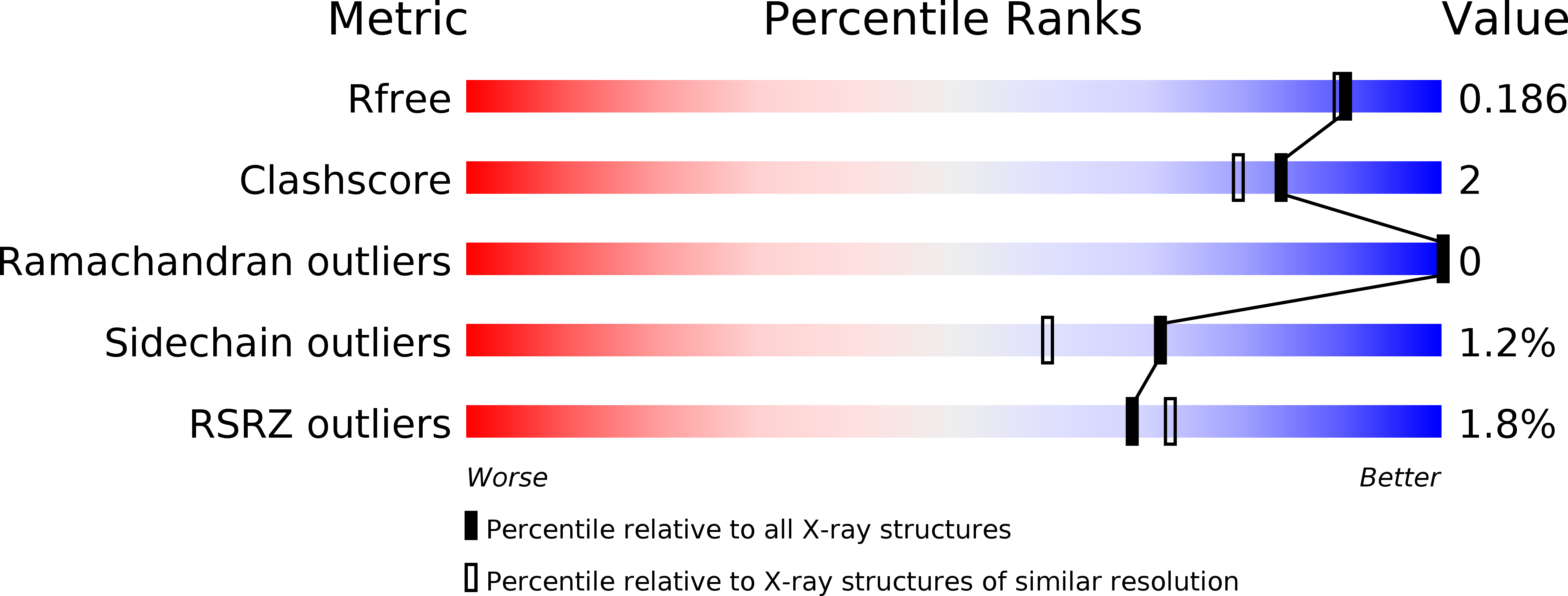
wwPDB Validation 3D Report Full Report
Entity ID: 1 | |||||
|---|---|---|---|---|---|
| Molecule | Chains | Sequence Length | Organism | Details | Image |
| DNA polymerase III subunit beta | 387 | Rickettsia conorii str. Malish 7 | Mutation(s): 0 Gene Names: dnaN, RC0583 EC: 2.7.7.7 | 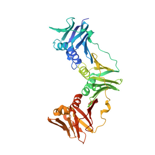 | |
UniProt | |||||
Find proteins for Q92I37 (Rickettsia conorii (strain ATCC VR-613 / Malish 7)) Explore Q92I37 Go to UniProtKB: Q92I37 | |||||
Entity Groups | |||||
| Sequence Clusters | 30% Identity50% Identity70% Identity90% Identity95% Identity100% Identity | ||||
| UniProt Group | Q92I37 | ||||
Sequence AnnotationsExpand | |||||
| |||||
| Ligands 2 Unique | |||||
|---|---|---|---|---|---|
| ID | Chains | Name / Formula / InChI Key | 2D Diagram | 3D Interactions | |
| MRD Query on MRD | E [auth A] | (4R)-2-METHYLPENTANE-2,4-DIOL C6 H14 O2 SVTBMSDMJJWYQN-RXMQYKEDSA-N |  | ||
| EDO Query on EDO | C [auth A], D [auth A], F [auth B] | 1,2-ETHANEDIOL C2 H6 O2 LYCAIKOWRPUZTN-UHFFFAOYSA-N |  | ||
| Length ( Å ) | Angle ( ˚ ) |
|---|---|
| a = 76.52 | α = 90 |
| b = 86.91 | β = 115.39 |
| c = 79.51 | γ = 90 |
| Software Name | Purpose |
|---|---|
| PHENIX | refinement |
| XSCALE | data scaling |
| PDB_EXTRACT | data extraction |
| XDS | data reduction |
| MoRDa | phasing |
| Coot | model building |
RCSB PDB (citation) is hosted by
RCSB PDB is a member of the

