


Crystal structure of polyubiquitin with 3 ub domains, domains 1 and 2, from Naegleria fowleri ATCC 30863
Abendroth, J., Mayclin, S.J., Lorimer, D.D., Edwards, T.E.To be published.
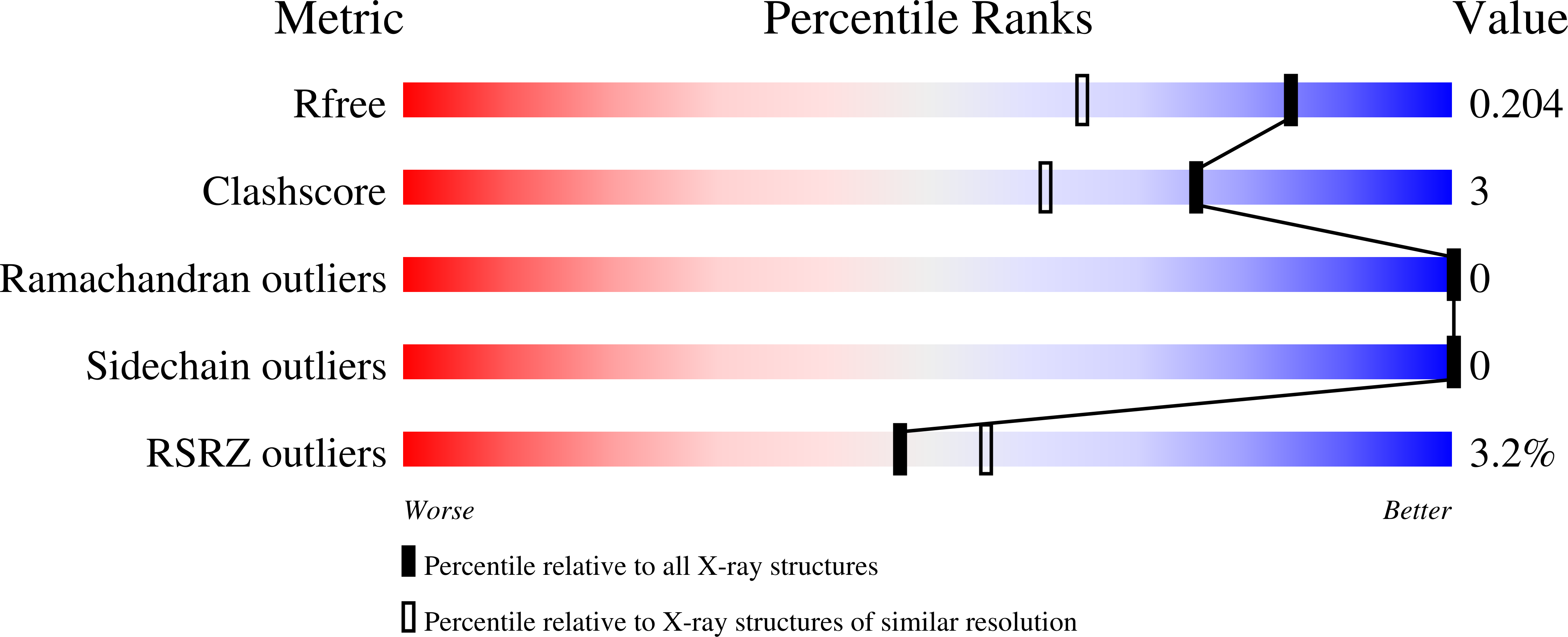
Experimental Data Snapshot
wwPDB Validation 3D Report Full Report
Entity ID: 1 | |||||
|---|---|---|---|---|---|
| Molecule | Chains | Sequence Length | Organism | Details | Image |
| Polyubiquitin | 160 | Naegleria fowleri | Mutation(s): 0 | 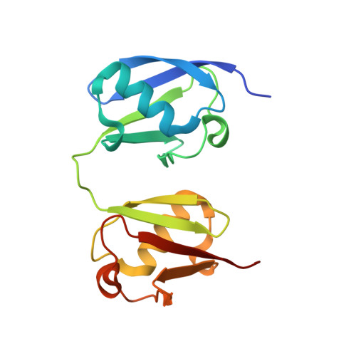 | |
UniProt | |||||
Find proteins for D2V6G7 (Naegleria gruberi) Explore D2V6G7 Go to UniProtKB: D2V6G7 | |||||
Entity Groups | |||||
| Sequence Clusters | 30% Identity50% Identity70% Identity90% Identity95% Identity100% Identity | ||||
| UniProt Group | D2V6G7 | ||||
Sequence AnnotationsExpand | |||||
| |||||
| Ligands 1 Unique | |||||
|---|---|---|---|---|---|
| ID | Chains | Name / Formula / InChI Key | 2D Diagram | 3D Interactions | |
| NO3 Query on NO3 | B [auth A], C [auth A], D [auth A] | NITRATE ION N O3 NHNBFGGVMKEFGY-UHFFFAOYSA-N |  | ||
| Length ( Å ) | Angle ( ˚ ) |
|---|---|
| a = 34.02 | α = 90 |
| b = 67.71 | β = 114.49 |
| c = 35.83 | γ = 90 |
| Software Name | Purpose |
|---|---|
| XSCALE | data scaling |
| PHENIX | refinement |
| PDB_EXTRACT | data extraction |
| XDS | data reduction |
| MOLREP | phasing |
| ARP | model building |
| Coot | model building |
RCSB PDB (citation) is hosted by
RCSB PDB is a member of the

