5VEN

Murine ectonucleotide pyrophosphatase / phosphodiesterase 5 (ENPP5, NPP5)


Experimental Data Snapshot

  • Method: X-RAY DIFFRACTION
  • Resolution: 1.69 Å
  • R-Value Free: 0.164 
  • R-Value Work: 0.129 
  • R-Value Observed: 0.130 

wwPDB Validation   3D Report Full Report


Ligand Structure Quality Assessment 


This is version 2.0 of the entry. See complete history


Literature

A key tyrosine substitution restricts nucleotide hydrolysis by the ectoenzyme NPP5.

Gorelik, A.Randriamihaja, A.Illes, K.Nagar, B.

(2017) FEBS J 284: 3718-3726

  • DOI: https://doi.org/10.1111/febs.14266
  • Primary Citation of Related Structures:  
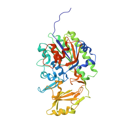
    5VEM, 5VEN, 5VEO

  • PubMed Abstract: 

    The ecto-nucleotide pyrophosphatase/phosphodiesterase (NPP) family of proteins mediates purinergic signaling by degrading extracellular nucleotides and also participates in phospholipid metabolism. NPP5 (ENPP5) is the least characterized member of this group and its specific role is unknown. This enzyme does not display activity on certain nucleotides and on other typical NPP substrates. In order to gain insights into its function, we determined the crystal structure of human and murine NPP5. Structural comparison with close homologs revealed a key phenylalanine to tyrosine substitution that prevents efficient hydrolysis of nucleotide diphosphates and triphosphates; reversal of this mutation enabled degradation of these molecules. Interestingly, NPP5 is able to cleave nicotinamide adenine dinucleotide (NAD), suggesting a potential role of this enzyme in NAD-based neurotransmission. An NPP5-specific metal binding motif is found adjacent to the active site, although its significance is unclear. These findings expand our understanding of substrate specificity within the NPP family.


  • Organizational Affiliation

    Department of Biochemistry and Groupe de Recherche Axé sur la Structure des Protéines, McGill University, Montreal, Canada.


Macromolecules
Find similar proteins by:  (by identity cutoff)  |  3D Structure
Entity ID: 1
MoleculeChains Sequence LengthOrganismDetailsImage
Ectonucleotide pyrophosphatase/phosphodiesterase family member 5
A, B
416Mus musculusMutation(s): 0 
Gene Names: Enpp5
EC: 3.1
UniProt & NIH Common Fund Data Resources
Find proteins for Q9EQG7 (Mus musculus)
Explore Q9EQG7 
Go to UniProtKB:  Q9EQG7
IMPC:  MGI:1933830
Entity Groups  
Sequence Clusters30% Identity50% Identity70% Identity90% Identity95% Identity100% Identity
UniProt GroupQ9EQG7
Sequence Annotations
Expand
  • Reference Sequence
Oligosaccharides

Help

Entity ID: 2
MoleculeChains Length2D Diagram Glycosylation3D Interactions
alpha-D-mannopyranose-(1-3)-[alpha-D-mannopyranose-(1-6)]beta-D-mannopyranose-(1-4)-2-acetamido-2-deoxy-beta-D-glucopyranose-(1-4)-2-acetamido-2-deoxy-beta-D-glucopyranose
C, G
5N-Glycosylation
Glycosylation Resources
GlyTouCan:  G22768VO
GlyCosmos:  G22768VO
GlyGen:  G22768VO
Entity ID: 3
MoleculeChains Length2D Diagram Glycosylation3D Interactions
2-acetamido-2-deoxy-beta-D-glucopyranose-(1-4)-2-acetamido-2-deoxy-beta-D-glucopyranose
D, E, H, I
2N-Glycosylation
Glycosylation Resources
GlyTouCan:  G42666HT
GlyCosmos:  G42666HT
GlyGen:  G42666HT
Entity ID: 4
MoleculeChains Length2D Diagram Glycosylation3D Interactions
alpha-L-fucopyranose-(1-6)-2-acetamido-2-deoxy-beta-D-glucopyranose
F, J
2N-Glycosylation
Glycosylation Resources
GlyTouCan:  G86851RC
GlyCosmos:  G86851RC
GlyGen:  G86851RC
Small Molecules
Ligands 3 Unique
IDChains Name / Formula / InChI Key2D Diagram3D Interactions
NAG
Query on NAG

Download Ideal Coordinates CCD File 
O [auth A],
P [auth A],
V [auth B],
W [auth B]
2-acetamido-2-deoxy-beta-D-glucopyranose
C8 H15 N O6
OVRNDRQMDRJTHS-FMDGEEDCSA-N
PG4
Query on PG4

Download Ideal Coordinates CCD File 
Q [auth A],
X [auth B]
TETRAETHYLENE GLYCOL
C8 H18 O5
UWHCKJMYHZGTIT-UHFFFAOYSA-N
ZN
Query on ZN

Download Ideal Coordinates CCD File 
K [auth A]
L [auth A]
M [auth A]
N [auth A]
R [auth B]
K [auth A],
L [auth A],
M [auth A],
N [auth A],
R [auth B],
S [auth B],
T [auth B],
U [auth B]
ZINC ION
Zn
PTFCDOFLOPIGGS-UHFFFAOYSA-N
Experimental Data & Validation

Experimental Data

  • Method: X-RAY DIFFRACTION
  • Resolution: 1.69 Å
  • R-Value Free: 0.164 
  • R-Value Work: 0.129 
  • R-Value Observed: 0.130 
  • Space Group: P 32 2 1
Unit Cell:
Length ( Å )Angle ( ˚ )
a = 82.403α = 90
b = 82.403β = 90
c = 301.01γ = 120
Software Package:
Software NamePurpose
PHENIXrefinement
HKL-2000data reduction
HKL-2000data scaling
PHASERphasing

Structure Validation

View Full Validation Report


Ligand Structure Quality Assessment 


Entry History & Funding Information

Deposition Data


Funding OrganizationLocationGrant Number
Canadian Institutes of Health Research (CIHR)CanadaMOP-133535

Revision History  (Full details and data files)

  • Version 1.0: 2017-09-20
    Type: Initial release
  • Version 1.1: 2017-11-22
    Changes: Database references
  • Version 1.2: 2020-01-08
    Changes: Author supporting evidence, Data collection
  • Version 2.0: 2020-07-29
    Type: Remediation
    Reason: Carbohydrate remediation
    Changes: Atomic model, Data collection, Derived calculations, Structure summary