5VDC

Crystal structure of the human DPF2 tandem PHD finger domain


Experimental Data Snapshot

  • Method: X-RAY DIFFRACTION
  • Resolution: 1.60 Å
  • R-Value Free: 0.191 
  • R-Value Work: 0.141 
  • R-Value Observed: 0.143 

wwPDB Validation   3D Report Full Report


This is version 1.5 of the entry. See complete history


Literature

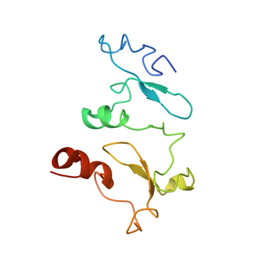
Histone-binding of DPF2 mediates its repressive role in myeloid differentiation.

Huber, F.M.Greenblatt, S.M.Davenport, A.M.Martinez, C.Xu, Y.Vu, L.P.Nimer, S.D.Hoelz, A.

(2017) Proc Natl Acad Sci U S A 114: 6016-6021

  • DOI: https://doi.org/10.1073/pnas.1700328114
  • Primary Citation of Related Structures:  
    5VDC

  • PubMed Abstract: 

    Double plant homeodomain finger 2 (DPF2) is a highly evolutionarily conserved member of the d4 protein family that is ubiquitously expressed in human tissues and was recently shown to inhibit the myeloid differentiation of hematopoietic stem/progenitor and acute myelogenous leukemia cells. Here, we present the crystal structure of the tandem plant homeodomain finger domain of human DPF2 at 1.6-Å resolution. We show that DPF2 interacts with the acetylated tails of both histones 3 and 4 via bipartite binding pockets on the DPF2 surface. Blocking these interactions through targeted mutagenesis of DPF2 abolishes its recruitment to target chromatin regions as well as its ability to prevent myeloid differentiation in vivo. Our findings suggest that the histone binding of DPF2 plays an important regulatory role in the transcriptional program that drives myeloid differentiation.


  • Organizational Affiliation

    Division of Chemistry and Chemical Engineering, California Institute of Technology, Pasadena, CA 91125.


Macromolecules
Find similar proteins by:  (by identity cutoff)  |  3D Structure
Entity ID: 1
MoleculeChains Sequence LengthOrganismDetailsImage
Zinc finger protein ubi-d4126Homo sapiensMutation(s): 0 
Gene Names: DPF2BAF45DREQUBID4
UniProt & NIH Common Fund Data Resources
Find proteins for Q92785 (Homo sapiens)
Explore Q92785 
Go to UniProtKB:  Q92785
PHAROS:  Q92785
GTEx:  ENSG00000133884 
Entity Groups  
Sequence Clusters30% Identity50% Identity70% Identity90% Identity95% Identity100% Identity
UniProt GroupQ92785
Sequence Annotations
Expand
  • Reference Sequence
Small Molecules
Ligands 3 Unique
IDChains Name / Formula / InChI Key2D Diagram3D Interactions
GOL
Query on GOL

Download Ideal Coordinates CCD File 
F [auth A],
G [auth A]
GLYCEROL
C3 H8 O3
PEDCQBHIVMGVHV-UHFFFAOYSA-N
ZN
Query on ZN

Download Ideal Coordinates CCD File 
B [auth A],
C [auth A],
D [auth A],
E [auth A]
ZINC ION
Zn
PTFCDOFLOPIGGS-UHFFFAOYSA-N
EDO
Query on EDO

Download Ideal Coordinates CCD File 
H [auth A],
I [auth A],
J [auth A]
1,2-ETHANEDIOL
C2 H6 O2
LYCAIKOWRPUZTN-UHFFFAOYSA-N
Experimental Data & Validation

Experimental Data

  • Method: X-RAY DIFFRACTION
  • Resolution: 1.60 Å
  • R-Value Free: 0.191 
  • R-Value Work: 0.141 
  • R-Value Observed: 0.143 
  • Space Group: I 4 2 2
Unit Cell:
Length ( Å )Angle ( ˚ )
a = 107.5α = 90
b = 107.5β = 90
c = 53.3γ = 90
Software Package:
Software NamePurpose
PHENIXrefinement
XSCALEdata scaling
AutoSolphasing
XDSdata reduction

Structure Validation

View Full Validation Report


Entry History & Funding Information

Deposition Data


Funding OrganizationLocationGrant Number
Heritage Medical Research InstituteUnited States--
Howard Hughes Medical Institute (HHMI)United States--

Revision History  (Full details and data files)

  • Version 1.0: 2017-05-24
    Type: Initial release
  • Version 1.1: 2017-06-07
    Changes: Database references
  • Version 1.2: 2017-06-21
    Changes: Database references
  • Version 1.3: 2017-09-27
    Changes: Author supporting evidence
  • Version 1.4: 2019-11-20
    Changes: Author supporting evidence
  • Version 1.5: 2024-03-06
    Changes: Data collection, Database references