5V00

Structure of HutD from Pseudomonas fluorescens SBW25 (Formate condition)


Experimental Data Snapshot

  • Method: X-RAY DIFFRACTION
  • Resolution: 1.80 Å
  • R-Value Free: 0.196 
  • R-Value Work: 0.167 
  • R-Value Observed: 0.168 

wwPDB Validation   3D Report Full Report


This is version 1.4 of the entry. See complete history


Literature

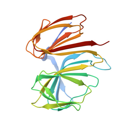
Crystal structure of a bicupin protein HutD involved in histidine utilization in Pseudomonas.

Gerth, M.L.Liu, Y.Jiao, W.Zhang, X.X.Baker, E.N.Lott, J.S.Rainey, P.B.Johnston, J.M.

(2017) Proteins 85: 1580-1588

  • DOI: https://doi.org/10.1002/prot.25303
  • Primary Citation of Related Structures:  
    5V00

  • PubMed Abstract: 

    Cupins form one of the most functionally diverse superfamilies of proteins, with members performing a wide range of catalytic, non-catalytic, and regulatory functions. HutD is a predicted bicupin protein that is involved in histidine utilization (Hut) in Pseudomonas species. Previous genetic analyses have suggested that it limits the upper level of Hut pathway expression, but its mechanism of action is unknown. Here, we have determined the structure of PfluHutD at 1.74 Å resolution in several crystallization conditions, and identified N-formyl-l-glutamate (FG, a Hut pathway intermediate) as a potential ligand in vivo. Proteins 2017; 85:1580-1588. © 2017 Wiley Periodicals, Inc.


  • Organizational Affiliation

    New Zealand Institute for Advanced Study, Massey University Albany, Auckland, New Zealand.


Macromolecules
Find similar proteins by:  (by identity cutoff)  |  3D Structure
Entity ID: 1
MoleculeChains Sequence LengthOrganismDetailsImage
Uncharacterized protein
A, B
193Pseudomonas fluorescens SBW25Mutation(s): 0 
Gene Names: PFLU_0360
UniProt
Find proteins for C3K802 (Pseudomonas fluorescens (strain SBW25))
Explore C3K802 
Go to UniProtKB:  C3K802
Entity Groups  
Sequence Clusters30% Identity50% Identity70% Identity90% Identity95% Identity100% Identity
UniProt GroupC3K802
Sequence Annotations
Expand
  • Reference Sequence
Small Molecules
Ligands 2 Unique
IDChains Name / Formula / InChI Key2D Diagram3D Interactions
GOL
Query on GOL

Download Ideal Coordinates CCD File 
C [auth A],
N [auth B]
GLYCEROL
C3 H8 O3
PEDCQBHIVMGVHV-UHFFFAOYSA-N
FMT
Query on FMT

Download Ideal Coordinates CCD File 
D [auth A]
E [auth A]
F [auth A]
G [auth A]
H [auth A]
D [auth A],
E [auth A],
F [auth A],
G [auth A],
H [auth A],
I [auth B],
J [auth B],
K [auth B],
L [auth B],
M [auth B]
FORMIC ACID
C H2 O2
BDAGIHXWWSANSR-UHFFFAOYSA-N
Experimental Data & Validation

Experimental Data

  • Method: X-RAY DIFFRACTION
  • Resolution: 1.80 Å
  • R-Value Free: 0.196 
  • R-Value Work: 0.167 
  • R-Value Observed: 0.168 
  • Space Group: P 41 21 2
Unit Cell:
Length ( Å )Angle ( ˚ )
a = 82.863α = 90
b = 82.863β = 90
c = 174.419γ = 90
Software Package:
Software NamePurpose
SCALEPACKdata scaling
REFMACrefinement
PDB_EXTRACTdata extraction
DENZOdata reduction
PHASERphasing

Structure Validation

View Full Validation Report


Entry History & Funding Information

Deposition Data


Funding OrganizationLocationGrant Number
Foundation for Research Science and TechnologyNew Zealand--
Health Research Council (HRC)New Zealand--

Revision History  (Full details and data files)

  • Version 1.0: 2017-03-15
    Type: Initial release
  • Version 1.1: 2017-04-19
    Changes: Database references
  • Version 1.2: 2017-08-02
    Changes: Database references
  • Version 1.3: 2020-01-01
    Changes: Author supporting evidence
  • Version 1.4: 2023-10-04
    Changes: Data collection, Database references, Refinement description