5U3U

Human PPARdelta ligand-binding domain in complexed with specific agonist 5


Experimental Data Snapshot

  • Method: X-RAY DIFFRACTION
  • Resolution: 2.10 Å
  • R-Value Free: 0.261 
  • R-Value Work: 0.207 
  • R-Value Observed: 0.209 

wwPDB Validation   3D Report Full Report


This is version 2.1 of the entry. See complete history


Literature

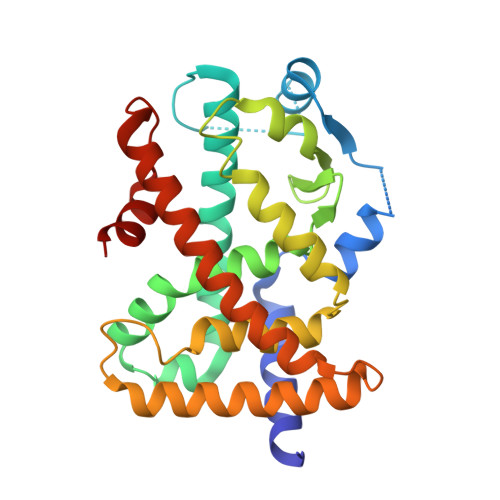
Structural basis for specific ligation of the peroxisome proliferator-activated receptor delta.

Wu, C.C.Baiga, T.J.Downes, M.La Clair, J.J.Atkins, A.R.Richard, S.B.Fan, W.Stockley-Noel, T.A.Bowman, M.E.Noel, J.P.Evans, R.M.

(2017) Proc Natl Acad Sci U S A 114: E2563-E2570

  • DOI: https://doi.org/10.1073/pnas.1621513114
  • Primary Citation of Related Structures:  
    5U3Q, 5U3R, 5U3S, 5U3T, 5U3U, 5U3V, 5U3W, 5U3X, 5U3Y, 5U3Z, 5U40, 5U41, 5U42, 5U43, 5U44, 5U45, 5U46

  • PubMed Abstract: 

    The peroxisome proliferator-activated receptor (PPAR) family comprises three subtypes: PPARα, PPARγ, and PPARδ. PPARδ transcriptionally modulates lipid metabolism and the control of energy homeostasis; therefore, PPARδ agonists are promising agents for treating a variety of metabolic disorders. In the present study, we develop a panel of rationally designed PPARδ agonists. The modular motif affords efficient syntheses using building blocks optimized for interactions with subtype-specific residues in the PPARδ ligand-binding domain (LBD). A combination of atomic-resolution protein X-ray crystallographic structures, ligand-dependent LBD stabilization assays, and cell-based transactivation measurements delineate structure-activity relationships (SARs) for PPARδ-selective targeting and structural modulation. We identify key ligand-induced conformational transitions of a conserved tryptophan side chain in the LBD that trigger reorganization of the H2'-H3 surface segment of PPARδ. The subtype-specific conservation of H2'-H3 sequences suggests that this architectural remodeling constitutes a previously unrecognized conformational switch accompanying ligand-dependent PPARδ transcriptional regulation.


  • Organizational Affiliation

    Howard Hughes Medical Institute, The Salk Institute for Biological Studies, La Jolla, CA 92037.


Macromolecules
Find similar proteins by:  (by identity cutoff)  |  3D Structure
Entity ID: 1
MoleculeChains Sequence LengthOrganismDetailsImage
Peroxisome proliferator-activated receptor delta
A, B
272Homo sapiensMutation(s): 0 
Gene Names: PPARDNR1C2PPARB
UniProt & NIH Common Fund Data Resources
Find proteins for Q03181 (Homo sapiens)
Explore Q03181 
Go to UniProtKB:  Q03181
PHAROS:  Q03181
GTEx:  ENSG00000112033 
Entity Groups  
Sequence Clusters30% Identity50% Identity70% Identity90% Identity95% Identity100% Identity
UniProt GroupQ03181
Sequence Annotations
Expand
  • Reference Sequence
Experimental Data & Validation

Experimental Data

  • Method: X-RAY DIFFRACTION
  • Resolution: 2.10 Å
  • R-Value Free: 0.261 
  • R-Value Work: 0.207 
  • R-Value Observed: 0.209 
  • Space Group: P 1 21 1
Unit Cell:
Length ( Å )Angle ( ˚ )
a = 39.6α = 90
b = 94.55β = 97.91
c = 96.57γ = 90
Software Package:
Software NamePurpose
PHENIXrefinement
HKL-2000data reduction
HKL-2000data scaling
PHASERphasing

Structure Validation

View Full Validation Report


Entry History 

Deposition Data

Revision History  (Full details and data files)

  • Version 1.0: 2017-03-22
    Type: Initial release
  • Version 1.1: 2017-03-29
    Changes: Database references
  • Version 1.2: 2017-04-05
    Changes: Database references
  • Version 2.0: 2020-07-29
    Type: Remediation
    Reason: Carbohydrate remediation
    Changes: Atomic model, Data collection, Derived calculations, Structure summary
  • Version 2.1: 2023-10-04
    Changes: Data collection, Database references, Refinement description, Structure summary