5T20

Crystal Structure of Tarin Lectin bound to Trimannose


Experimental Data Snapshot

  • Method: X-RAY DIFFRACTION
  • Resolution: 1.91 Å
  • R-Value Free: 0.244 
  • R-Value Work: 0.209 
  • R-Value Observed: 0.211 

wwPDB Validation   3D Report Full Report


Ligand Structure Quality Assessment 


This is version 2.1 of the entry. See complete history


Literature

High-resolution crystal structures of Colocasia esculenta tarin lectin.

Pereira, P.R.Meagher, J.L.Winter, H.C.Goldstein, I.J.Paschoalin, V.M.Silva, J.T.Stuckey, J.A.

(2017) Glycobiology 27: 50-56

  • DOI: https://doi.org/10.1093/glycob/cww083
  • Primary Citation of Related Structures:  
    5T1X, 5T20

  • PubMed Abstract: 

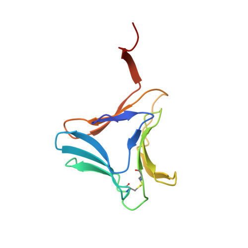
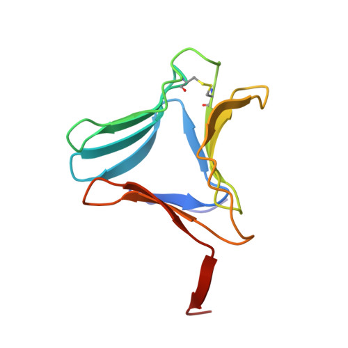
    Tarin, the Colocasia esculenta lectin from the superfamily of α-d-mannose-specific plant bulb lectins, is a tetramer of 47 kDa composed of two heterodimers. Each heterodimer possesses homologous monomers of ~11.9 (A chain) and ~12.7 (B chain) kDa. The structures of apo and carbohydrate-bound tarin were solved to 1.7 Å and 1.91 Å, respectively. Each tarin monomer forms a canonical β-prism II fold, common to all members of Galanthus nivalis agglutinin (GNA) family, which is partially stabilized by a disulfide bond and a conserved hydrophobic core. The heterodimer is formed through domain swapping involving the C-terminal β-strand and the β-sheet on face I of the prism. The tetramer is assembled through the dimerization of the B chains from heterodimers involving face II of each prism. The 1.91 Å crystal structure of tarin bound to Manα(1,3)Manα(1,6)Man reveals an expanded carbohydrate-binding sequence (QxDxNxVxYx 4/6 WX) on face III of the β-prism. Both monomers possess a similar fold, except for the length of the loop, which begins after the conserved tyrosine and creates the binding pocket for the α(1,6)-terminal mannose. This loop differs in size and amino-acid composition from 10 other β-prism II domain proteins, and may confer carbohydrate-binding specificity among members of the GNA-related lectin family.


  • Organizational Affiliation

    Centro de Tecnologia, Universidade Federal do Rio de Janeiro (UFRJ), Avenida Athos da Silveira Ramos, 149., Rio de Janeiro 21941-909, Brazil.


Macromolecules
Find similar proteins by:  (by identity cutoff)  |  3D Structure
Entity ID: 1
MoleculeChains Sequence LengthOrganismDetailsImage
Lectin
A, C, E, G, I
A, C, E, G, I, K, M, O
110Colocasia esculentaMutation(s): 0 
UniProt
Find proteins for A5HMM7 (Colocasia esculenta)
Explore A5HMM7 
Go to UniProtKB:  A5HMM7
Entity Groups  
Sequence Clusters30% Identity50% Identity70% Identity90% Identity95% Identity100% Identity
UniProt GroupA5HMM7
Sequence Annotations
Expand
  • Reference Sequence
Find similar proteins by:  (by identity cutoff)  |  3D Structure
Entity ID: 2
MoleculeChains Sequence LengthOrganismDetailsImage
Lectin
B, D, F, H, J
B, D, F, H, J, L, N, P
110Colocasia esculentaMutation(s): 0 
UniProt
Find proteins for Q39487 (Colocasia esculenta)
Explore Q39487 
Go to UniProtKB:  Q39487
Entity Groups  
Sequence Clusters30% Identity50% Identity70% Identity90% Identity95% Identity100% Identity
UniProt GroupQ39487
Sequence Annotations
Expand
  • Reference Sequence
Oligosaccharides

Help

Entity ID: 4
MoleculeChains Length2D Diagram Glycosylation3D Interactions
alpha-D-mannopyranose-(1-6)-alpha-D-mannopyranose
V
2N/A
Glycosylation Resources
GlyTouCan:  G77384RA
GlyCosmos:  G77384RA
GlyGen:  G77384RA
Biologically Interesting Molecules (External Reference) 1 Unique
Experimental Data & Validation

Experimental Data

  • Method: X-RAY DIFFRACTION
  • Resolution: 1.91 Å
  • R-Value Free: 0.244 
  • R-Value Work: 0.209 
  • R-Value Observed: 0.211 
  • Space Group: P 1
Unit Cell:
Length ( Å )Angle ( ˚ )
a = 78.28α = 76.19
b = 78.828β = 70.44
c = 92.935γ = 59.8
Software Package:
Software NamePurpose
HKL-2000data reduction
PDB_EXTRACTdata extraction
HKL-2000data scaling
BUSTERrefinement
PHASERphasing
DENZOdata reduction
SCALEPACKdata scaling

Structure Validation

View Full Validation Report


Ligand Structure Quality Assessment 


Entry History 

Deposition Data

Revision History  (Full details and data files)

  • Version 1.0: 2016-09-14
    Type: Initial release
  • Version 1.1: 2017-01-11
    Changes: Database references
  • Version 1.2: 2017-11-22
    Changes: Advisory, Refinement description
  • Version 2.0: 2020-07-29
    Type: Remediation
    Reason: Carbohydrate remediation
    Changes: Advisory, Atomic model, Data collection, Derived calculations, Structure summary
  • Version 2.1: 2023-10-04
    Changes: Data collection, Database references, Refinement description, Structure summary