5SVO

Structure of IDH2 mutant R140Q


Experimental Data Snapshot

  • Method: X-RAY DIFFRACTION
  • Resolution: 1.87 Å
  • R-Value Free: 0.218 
  • R-Value Work: 0.190 
  • R-Value Observed: 0.191 

wwPDB Validation   3D Report Full Report


Ligand Structure Quality Assessment 


This is version 1.2 of the entry. See complete history


Literature

Allosteric Mutant IDH1 Inhibitors Reveal Mechanisms for IDH1 Mutant and Isoform Selectivity.

Xie, X.Baird, D.Bowen, K.Capka, V.Chen, J.Chenail, G.Cho, Y.Dooley, J.Farsidjani, A.Fortin, P.Kohls, D.Kulathila, R.Lin, F.McKay, D.Rodrigues, L.Sage, D.Toure, B.B.van der Plas, S.Wright, K.Xu, M.Yin, H.Levell, J.Pagliarini, R.A.

(2017) Structure 25: 506-513

  • DOI: https://doi.org/10.1016/j.str.2016.12.017
  • Primary Citation of Related Structures:  
    5SUN, 5SVF, 5SVN, 5SVO

  • PubMed Abstract: 

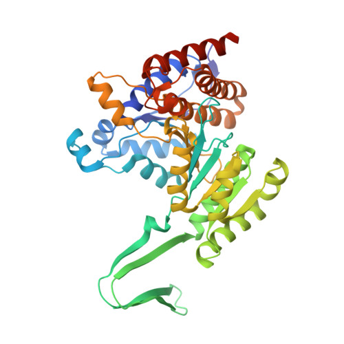
    Oncogenic IDH1 and IDH2 mutations contribute to cancer via production of R-2-hydroxyglutarate (2-HG). Here, we characterize two structurally distinct mutant- and isoform-selective IDH1 inhibitors that inhibit 2-HG production. Both bind to an allosteric pocket on IDH1, yet shape it differently, highlighting the plasticity of this site. Oncogenic IDH1 R132H mutation destabilizes an IDH1 "regulatory segment," which otherwise restricts compound access to the allosteric pocket. Regulatory segment destabilization in wild-type IDH1 promotes inhibitor binding, suggesting that destabilization is critical for mutant selectivity. We also report crystal structures of oncogenic IDH2 mutant isoforms, highlighting the fact that the analogous segment of IDH2 is not similarly destabilized. This intrinsic stability of IDH2 may contribute to observed inhibitor IDH1 isoform selectivity. Moreover, discrete residues in the IDH1 allosteric pocket that differ from IDH2 may also guide IDH1 isoform selectivity. These data provide a deeper understanding of how IDH1 inhibitors achieve mutant and isoform selectivity.


  • Organizational Affiliation

    Center for Proteomic Chemistry, Novartis Institutes for Biomedical Research, 250 Massachusetts Avenue, Cambridge, MA 02139, USA.


Macromolecules
Find similar proteins by:  (by identity cutoff)  |  3D Structure
Entity ID: 1
MoleculeChains Sequence LengthOrganismDetailsImage
Isocitrate dehydrogenase [NADP], mitochondrial
A, B
424Homo sapiensMutation(s): 1 
Gene Names: IDH2
EC: 1.1.1.42
UniProt & NIH Common Fund Data Resources
Find proteins for P48735 (Homo sapiens)
Explore P48735 
Go to UniProtKB:  P48735
PHAROS:  P48735
GTEx:  ENSG00000182054 
Entity Groups  
Sequence Clusters30% Identity50% Identity70% Identity90% Identity95% Identity100% Identity
UniProt GroupP48735
Sequence Annotations
Expand
  • Reference Sequence
Small Molecules
Ligands 2 Unique
IDChains Name / Formula / InChI Key2D Diagram3D Interactions
NAP
Query on NAP

Download Ideal Coordinates CCD File 
C [auth A],
E [auth B]
NADP NICOTINAMIDE-ADENINE-DINUCLEOTIDE PHOSPHATE
C21 H28 N7 O17 P3
XJLXINKUBYWONI-NNYOXOHSSA-N
EPE
Query on EPE

Download Ideal Coordinates CCD File 
D [auth A]4-(2-HYDROXYETHYL)-1-PIPERAZINE ETHANESULFONIC ACID
C8 H18 N2 O4 S
JKMHFZQWWAIEOD-UHFFFAOYSA-N
Experimental Data & Validation

Experimental Data

  • Method: X-RAY DIFFRACTION
  • Resolution: 1.87 Å
  • R-Value Free: 0.218 
  • R-Value Work: 0.190 
  • R-Value Observed: 0.191 
  • Space Group: P 21 21 21
Unit Cell:
Length ( Å )Angle ( ˚ )
a = 58.661α = 90
b = 119.207β = 90
c = 127.496γ = 90
Software Package:
Software NamePurpose
BUSTERrefinement
XDSdata reduction
autoPROCdata scaling
PHASERphasing

Structure Validation

View Full Validation Report


Ligand Structure Quality Assessment 


Entry History 

Deposition Data

Revision History  (Full details and data files)

  • Version 1.0: 2017-02-08
    Type: Initial release
  • Version 1.1: 2017-03-22
    Changes: Database references
  • Version 1.2: 2024-03-06
    Changes: Data collection, Database references, Refinement description