5OV4

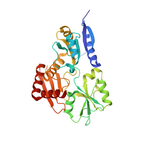
Bacillus megaterium porphobilinogen deaminase D82A mutant


Experimental Data Snapshot

  • Method: X-RAY DIFFRACTION
  • Resolution: 2.69 Å
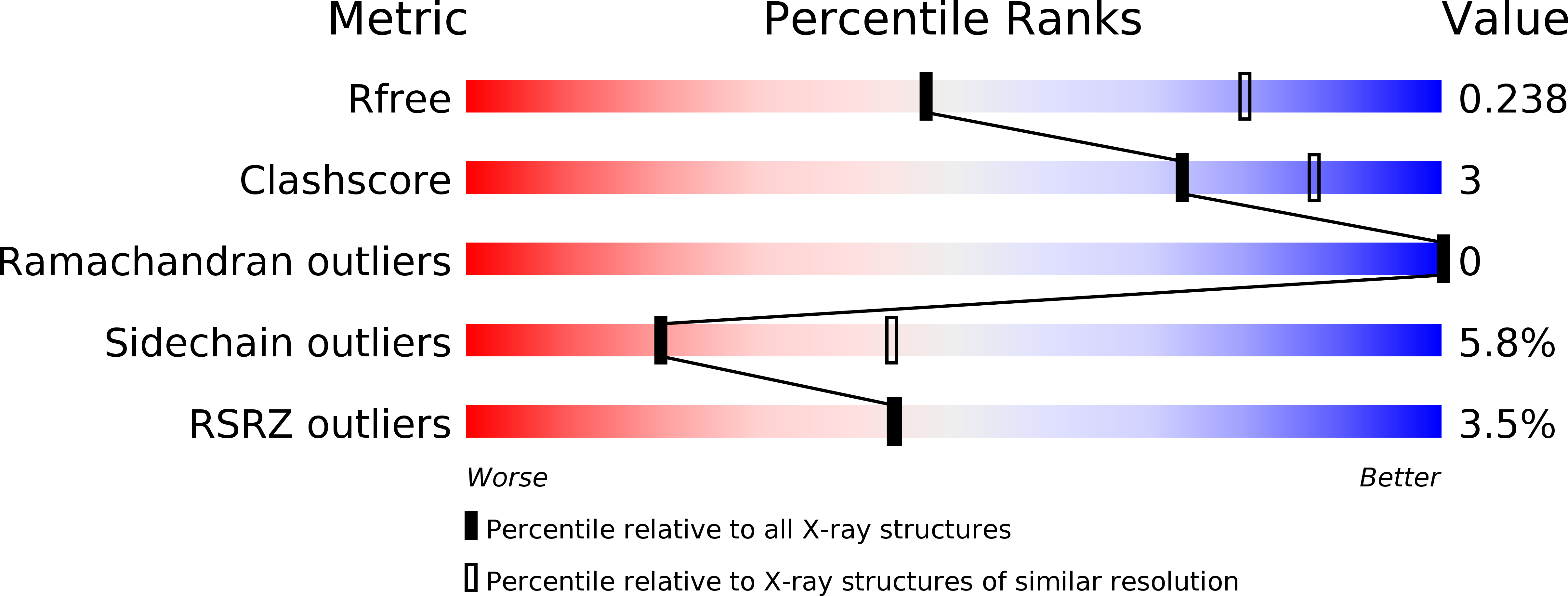
  • R-Value Free: 0.239 
  • R-Value Work: 0.209 
  • R-Value Observed: 0.211 

wwPDB Validation   3D Report Full Report


This is version 1.3 of the entry. See complete history


Literature

Structural studies of domain movement in active-site mutants of porphobilinogen deaminase from Bacillus megaterium.

Guo, J.Erskine, P.Coker, A.R.Wood, S.P.Cooper, J.B.

(2017) Acta Crystallogr F Struct Biol Commun 73: 612-620

  • DOI: https://doi.org/10.1107/S2053230X17015436
  • Primary Citation of Related Structures:  
    5OV4, 5OV5, 5OV6

  • PubMed Abstract: 

    The enzyme porphobilinogen deaminase (PBGD) is one of the key enzymes in tetrapyrrole biosynthesis. It catalyses the formation of a linear tetrapyrrole from four molecules of the substrate porphobilinogen (PBG). It has a dipyrromethane cofactor (DPM) in the active site which is covalently linked to a conserved cysteine residue through a thioether bridge. The substrate molecules are linked to the cofactor in a stepwise head-to-tail manner during the reaction, which is catalysed by a conserved aspartate residue: Asp82 in the B. megaterium enzyme. Three mutations have been made affecting Asp82 (D82A, D82E and D82N) and their crystal structures have been determined at resolutions of 2.7, 1.8 and 1.9 Å, respectively. These structures reveal that whilst the D82E mutant possesses the DPM cofactor, in the D82N and D82A mutants the cofactor is likely to be missing, incompletely assembled or disordered. Comparison of the mutant PBGD structures with that of the wild-type enzyme shows that there are significant domain movements and suggests that the enzyme adopts `open' and `closed' conformations, potentially in response to substrate binding.


  • Organizational Affiliation

    Division of Medicine, University College London, Gower Street, London WC1E 6BT, England.


Macromolecules
Find similar proteins by:  (by identity cutoff)  |  3D Structure
Entity ID: 1
MoleculeChains Sequence LengthOrganismDetailsImage
Porphobilinogen deaminase311Priestia megateriumMutation(s): 1 
Gene Names: hemC
EC: 2.5.1.61
UniProt
Find proteins for Q8GCA8 (Priestia megaterium)
Explore Q8GCA8 
Go to UniProtKB:  Q8GCA8
Entity Groups  
Sequence Clusters30% Identity50% Identity70% Identity90% Identity95% Identity100% Identity
UniProt GroupQ8GCA8
Sequence Annotations
Expand
  • Reference Sequence
Experimental Data & Validation

Experimental Data

  • Method: X-RAY DIFFRACTION
  • Resolution: 2.69 Å
  • R-Value Free: 0.239 
  • R-Value Work: 0.209 
  • R-Value Observed: 0.211 
  • Space Group: P 21 21 21
Unit Cell:
Length ( Å )Angle ( ˚ )
a = 49.092α = 90
b = 62.487β = 90
c = 91.391γ = 90
Software Package:
Software NamePurpose
PHENIXrefinement
REFMACrefinement
xia2data scaling
MOLREPphasing
Cootmodel building
XDSdata reduction

Structure Validation

View Full Validation Report


Entry History 

Deposition Data

Revision History  (Full details and data files)

  • Version 1.0: 2017-10-11
    Type: Initial release
  • Version 1.1: 2017-11-08
    Changes: Database references
  • Version 1.2: 2017-11-22
    Changes: Database references
  • Version 1.3: 2024-01-17
    Changes: Data collection, Database references, Refinement description