Role of SH3b binding domain in a natural deletion mutant of Kayvirus endolysin LysF1 with a broad range of lytic activity.
Benesik, M., Novacek, J., Janda, L., Dopitova, R., Pernisova, M., Melkova, K., Tisakova, L., Doskar, J., Zidek, L., Hejatko, J., Pantucek, R.(2018) Virus Genes 54: 130-139
- PubMed: 28852930
- DOI: https://doi.org/10.1007/s11262-017-1507-2
- Primary Citation of Related Structures:
5O1Q - PubMed Abstract:
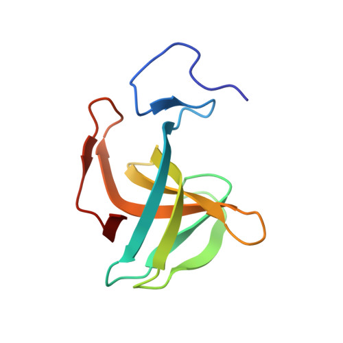
The spontaneous host-range mutants 812F1 and K1/420 are derived from polyvalent phage 812 that is almost identical to phage K, belonging to family Myoviridae and genus Kayvirus. Phage K1/420 is used for the phage therapy of staphylococcal infections. Endolysin of these mutants designated LysF1, consisting of an N-terminal cysteine-histidine-dependent aminohydrolase/peptidase (CHAP) domain and C-terminal SH3b cell wall-binding domain, has deleted middle amidase domain compared to wild-type endolysin. In this work, LysF1 and both its domains were prepared as recombinant proteins and their function was analyzed. LysF1 had an antimicrobial effect on 31 Staphylococcus species of the 43 tested. SH3b domain influenced antimicrobial activity of LysF1, since the lytic activity of the truncated variant containing the CHAP domain alone was decreased. The results of a co-sedimentation assay of SH3b domain showed that it was able to bind to three types of purified staphylococcal peptidoglycan 11.2, 11.3, and 11.8 that differ in their peptide bridge, but also to the peptidoglycan type 11.5 of Streptococcus uberis, and this capability was verified in vivo using the fusion protein with GFP and fluorescence microscopy. Using several different approaches, including NMR, we have not confirmed the previously proposed interaction of the SH3b domain with the pentaglycine bridge in the bacterial cell wall. The new naturally raised deletion mutant endolysin LysF1 is smaller than LysK, has a broad lytic spectrum, and therefore is an appropriate enzyme for practical use. The binding spectrum of SH3b domain covering all known staphylococcal peptidoglycan types is a promising feature for creating new chimeolysins by combining it with more effective catalytic domains.
Organizational Affiliation:
Department of Experimental Biology, Faculty of Science, Masaryk University, Kotlářská 2, Brno, 611 37, Czech Republic.