5NUR

Structural basis for maintenance of bacterial outer membrane lipid asymmetry


Experimental Data Snapshot

  • Method: X-RAY DIFFRACTION
  • Resolution: 3.29 Å
  • R-Value Free: 0.259 
  • R-Value Work: 0.219 
  • R-Value Observed: 0.221 

wwPDB Validation   3D Report Full Report


This is version 2.1 of the entry. See complete history


Literature

Structural basis for maintenance of bacterial outer membrane lipid asymmetry.

Abellon-Ruiz, J.Kaptan, S.S.Basle, A.Claudi, B.Bumann, D.Kleinekathofer, U.van den Berg, B.

(2017) Nat Microbiol 2: 1616-1623

  • DOI: https://doi.org/10.1038/s41564-017-0046-x
  • Primary Citation of Related Structures:  
    5NUO, 5NUP, 5NUQ, 5NUR

  • PubMed Abstract: 

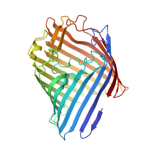
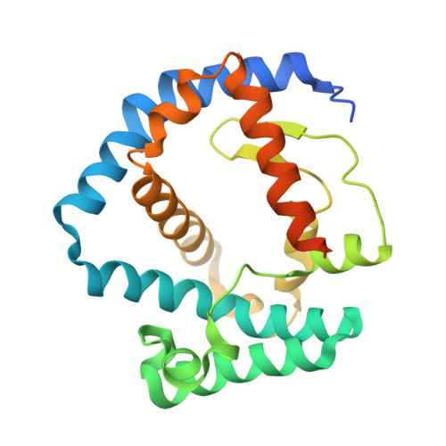
    The Gram-negative bacterial outer membrane (OM) is a unique bilayer that forms an efficient permeation barrier to protect the cell from noxious compounds 1 , 2 . The defining characteristic of the OM is lipid asymmetry, with phospholipids comprising the inner leaflet and lipopolysaccharides comprising the outer leaflet 1-3 . This asymmetry is maintained by the Mla pathway, a six-component system that is widespread in Gram-negative bacteria and is thought to mediate retrograde transport of misplaced phospholipids from the outer leaflet of the OM to the cytoplasmic membrane 4 . The OM lipoprotein MlaA performs the first step in this process via an unknown mechanism that does not require external energy input. Here we show, using X-ray crystallography, molecular dynamics simulations and in vitro and in vivo functional assays, that MlaA is a monomeric α-helical OM protein that functions as a phospholipid translocation channel, forming a ~20-Å-thick doughnut embedded in the inner leaflet of the OM with a central, amphipathic pore. This architecture prevents access of inner leaflet phospholipids to the pore, but allows outer leaflet phospholipids to bind to a pronounced ridge surrounding the channel, followed by diffusion towards the periplasmic space. Enterobacterial MlaA proteins form stable complexes with OmpF/C 5,6 , but the porins do not appear to play an active role in phospholipid transport. MlaA represents a lipid transport protein that selectively removes outer leaflet phospholipids to help maintain the essential barrier function of the bacterial OM.


  • Organizational Affiliation

    Institute for Cell and Molecular Biosciences, The Medical School, Newcastle University, Newcastle upon Tyne, NE2 4HH, UK.


Macromolecules
Find similar proteins by:  (by identity cutoff)  |  3D Structure
Entity ID: 1
MoleculeChains Sequence LengthOrganismDetailsImage
Outer membrane protein FA [auth C],
C [auth A],
E
340Escherichia coli K-12Mutation(s): 0 
Gene Names: ompFcmlBcoacrytolFb0929JW0912
Membrane Entity: Yes 
UniProt
Find proteins for P02931 (Escherichia coli (strain K12))
Explore P02931 
Go to UniProtKB:  P02931
Entity Groups  
Sequence Clusters30% Identity50% Identity70% Identity90% Identity95% Identity100% Identity
UniProt GroupP02931
Sequence Annotations
Expand
  • Reference Sequence
Find similar proteins by:  (by identity cutoff)  |  3D Structure
Entity ID: 2
MoleculeChains Sequence LengthOrganismDetailsImage
ABC transporter permeaseB [auth D],
D [auth B],
F
236Klebsiella pneumoniaeMutation(s): 0 
Gene Names: 
UniProt
Find proteins for A0A0W8AQT6 (Klebsiella pneumoniae)
Explore A0A0W8AQT6 
Go to UniProtKB:  A0A0W8AQT6
Entity Groups  
Sequence Clusters30% Identity50% Identity70% Identity90% Identity95% Identity100% Identity
UniProt GroupA0A0W8AQT6
Sequence Annotations
Expand
  • Reference Sequence
Oligosaccharides

Help

Entity ID: 3
MoleculeChains Length2D Diagram Glycosylation3D Interactions
3-deoxy-alpha-D-manno-oct-2-ulopyranosonic acid-(2-4)-3-deoxy-alpha-D-manno-oct-2-ulopyranosonic acid-(2-6)-2-amino-2-deoxy-4-O-phosphono-beta-D-glucopyranose-(1-6)-2-amino-2-deoxy-1-O-phosphono-alpha-D-glucopyranose
G
4N/A
Entity ID: 4
MoleculeChains Length2D Diagram Glycosylation3D Interactions
3-deoxy-alpha-D-manno-oct-2-ulopyranosonic acid-(3-4)-3-deoxy-alpha-D-manno-oct-2-ulopyranosonic acid-(2-6)-2-amino-2-deoxy-4-O-phosphono-beta-D-glucopyranose-(1-6)-2-amino-2-deoxy-1-O-phosphono-alpha-D-glucopyranose
H
4N/A
Entity ID: 5
MoleculeChains Length2D Diagram Glycosylation3D Interactions
3-deoxy-alpha-D-manno-oct-2-ulopyranosonic acid-(3-6)-2-amino-2-deoxy-4-O-phosphono-beta-D-glucopyranose-(1-6)-2-amino-2-deoxy-1-O-phosphono-alpha-D-glucopyranose
I
3N/A
Small Molecules
Ligands 3 Unique
IDChains Name / Formula / InChI Key2D Diagram3D Interactions
C8E
Query on C8E

Download Ideal Coordinates CCD File 
N [auth A],
O [auth A],
S [auth E]
(HYDROXYETHYLOXY)TRI(ETHYLOXY)OCTANE
C16 H34 O5
FEOZZFHAVXYAMB-UHFFFAOYSA-N
SO4
Query on SO4

Download Ideal Coordinates CCD File 
K [auth C]
L [auth C]
M [auth C]
Q [auth A]
R [auth A]
K [auth C],
L [auth C],
M [auth C],
Q [auth A],
R [auth A],
U [auth E],
V [auth E],
W [auth E]
SULFATE ION
O4 S
QAOWNCQODCNURD-UHFFFAOYSA-L
CA
Query on CA

Download Ideal Coordinates CCD File 
J [auth C],
P [auth A],
T [auth E]
CALCIUM ION
Ca
BHPQYMZQTOCNFJ-UHFFFAOYSA-N
Experimental Data & Validation

Experimental Data

  • Method: X-RAY DIFFRACTION
  • Resolution: 3.29 Å
  • R-Value Free: 0.259 
  • R-Value Work: 0.219 
  • R-Value Observed: 0.221 
  • Space Group: C 1 2 1
Unit Cell:
Length ( Å )Angle ( ˚ )
a = 190.55α = 90
b = 179.66β = 116.55
c = 133.39γ = 90
Software Package:
Software NamePurpose
XDSdata reduction
xia2data reduction
Aimlessdata scaling
PHASERphasing
PHENIXrefinement
Cootmodel building

Structure Validation

View Full Validation Report


Entry History & Funding Information

Deposition Data


Funding OrganizationLocationGrant Number
European UnionUnited Kingdom115525

Revision History  (Full details and data files)

  • Version 1.0: 2017-10-25
    Type: Initial release
  • Version 1.1: 2017-11-01
    Changes: Database references
  • Version 1.2: 2017-12-06
    Changes: Database references
  • Version 2.0: 2020-07-29
    Type: Remediation
    Reason: Carbohydrate remediation
    Changes: Advisory, Atomic model, Data collection, Derived calculations, Non-polymer description, Structure summary
  • Version 2.1: 2024-01-31
    Changes: Data collection, Database references, Refinement description