5NDB

Crystal structure of metallo-beta-lactamase SPM-1 complexed with cyclobutanone inhibitor


Experimental Data Snapshot

  • Method: X-RAY DIFFRACTION
  • Resolution: 2.38 Å
  • R-Value Free: 0.218 
  • R-Value Work: 0.179 
  • R-Value Observed: 0.181 

wwPDB Validation   3D Report Full Report


Ligand Structure Quality Assessment 


This is version 1.2 of the entry. See complete history


Literature

Cyclobutanone Mimics of Intermediates in Metallo-beta-Lactamase Catalysis.

Abboud, M.I.Kosmopoulou, M.Krismanich, A.P.Johnson, J.W.Hinchliffe, P.Brem, J.Claridge, T.D.W.Spencer, J.Schofield, C.J.Dmitrienko, G.I.

(2018) Chemistry 24: 5734-5737

  • DOI: https://doi.org/10.1002/chem.201705886
  • Primary Citation of Related Structures:  
    5NDB, 5NDE

  • PubMed Abstract: 

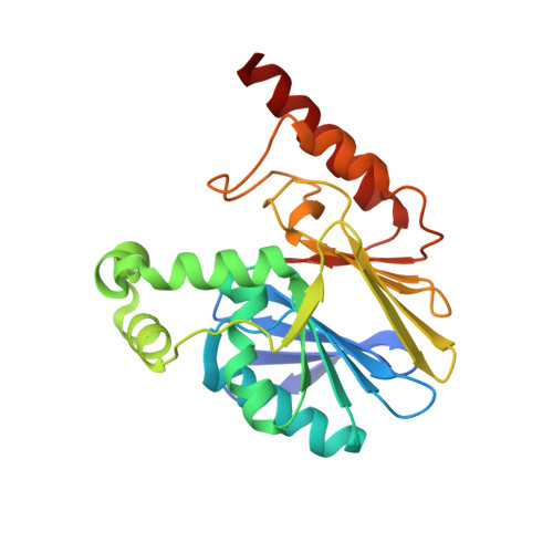
    The most important resistance mechanism to β-lactam antibiotics involves hydrolysis by two β-lactamase categories: the nucleophilic serine and the metallo-β-lactamases (SBLs and MBLs, respectively). Cyclobutanones are hydrolytically stable β-lactam analogues with potential to inhibit both SBLs and MBLs. We describe solution and crystallographic studies on the interaction of a cyclobutanone penem analogue with the clinically important MBL SPM-1. NMR experiments using 19 F-labeled SPM-1 imply the cyclobutanone binds to SPM-1 with micromolar affinity. A crystal structure of the SPM-1:cyclobutanone complex reveals binding of the hydrated cyclobutanone through interactions with one of the zinc ions, stabilisation of the hydrate by hydrogen bonding to zinc-bound water, and hydrophobic contacts with aromatic residues. NMR analyses using a 13 C-labeled cyclobutanone support assignment of the bound species as the hydrated ketone. The results inform on how MBLs bind substrates and stabilize tetrahedral intermediates. They support further investigations on the use of transition-state and/or intermediate analogues as inhibitors of all β-lactamase classes.


  • Organizational Affiliation

    Department of Chemistry, University of Oxford, 12 Mansfield Road, Oxford, OX1 3TA, UK.


Macromolecules
Find similar proteins by:  (by identity cutoff)  |  3D Structure
Entity ID: 1
MoleculeChains Sequence LengthOrganismDetailsImage
Beta-lactamase IMP-1
A, B
250Pseudomonas aeruginosaMutation(s): 0 
Gene Names: spm-1bla SPM-1blaSPM-1CCBH4851_00081ICEPaeSP_00028
EC: 3.5.2.6
UniProt
Find proteins for Q8G9Q0 (Pseudomonas aeruginosa)
Explore Q8G9Q0 
Go to UniProtKB:  Q8G9Q0
Entity Groups  
Sequence Clusters30% Identity50% Identity70% Identity90% Identity95% Identity100% Identity
UniProt GroupQ8G9Q0
Sequence Annotations
Expand
  • Reference Sequence
Small Molecules
Ligands 7 Unique
IDChains Name / Formula / InChI Key2D Diagram3D Interactions
8TW
Query on 8TW

Download Ideal Coordinates CCD File 
N [auth A],
W [auth B]
(1~{S},4~{R},5~{S})-7,7-bis(chloranyl)-6,6-bis(oxidanyl)-2$l^{4}-thiabicyclo[3.2.0]hept-2-ene-4-carboxylic acid
C7 H8 Cl2 O4 S
CYWQDVFNRSXKKZ-JJYYJPOSSA-N
TRS
Query on TRS

Download Ideal Coordinates CCD File 
Y [auth B]2-AMINO-2-HYDROXYMETHYL-PROPANE-1,3-DIOL
C4 H12 N O3
LENZDBCJOHFCAS-UHFFFAOYSA-O
SO4
Query on SO4

Download Ideal Coordinates CCD File 
X [auth B]SULFATE ION
O4 S
QAOWNCQODCNURD-UHFFFAOYSA-L
GOL
Query on GOL

Download Ideal Coordinates CCD File 
E [auth A]
F [auth A]
G [auth A]
H [auth A]
Q [auth B]
E [auth A],
F [auth A],
G [auth A],
H [auth A],
Q [auth B],
R [auth B],
S [auth B]
GLYCEROL
C3 H8 O3
PEDCQBHIVMGVHV-UHFFFAOYSA-N
DMS
Query on DMS

Download Ideal Coordinates CCD File 
J [auth A]DIMETHYL SULFOXIDE
C2 H6 O S
IAZDPXIOMUYVGZ-UHFFFAOYSA-N
ZN
Query on ZN

Download Ideal Coordinates CCD File 
C [auth A]
D [auth A]
I [auth A]
O [auth B]
P [auth B]
C [auth A],
D [auth A],
I [auth A],
O [auth B],
P [auth B],
T [auth B]
ZINC ION
Zn
PTFCDOFLOPIGGS-UHFFFAOYSA-N
CL
Query on CL

Download Ideal Coordinates CCD File 
K [auth A],
L [auth A],
M [auth A],
U [auth B],
V [auth B]
CHLORIDE ION
Cl
VEXZGXHMUGYJMC-UHFFFAOYSA-M
Experimental Data & Validation

Experimental Data

  • Method: X-RAY DIFFRACTION
  • Resolution: 2.38 Å
  • R-Value Free: 0.218 
  • R-Value Work: 0.179 
  • R-Value Observed: 0.181 
  • Space Group: P 42 2 2
Unit Cell:
Length ( Å )Angle ( ˚ )
a = 130.758α = 90
b = 130.758β = 90
c = 102.759γ = 90
Software Package:
Software NamePurpose
PHENIXrefinement
XDSdata reduction
Aimlessdata scaling
PHASERphasing

Structure Validation

View Full Validation Report


Ligand Structure Quality Assessment 


Entry History & Funding Information

Deposition Data


Funding OrganizationLocationGrant Number
Medical Research Council (United Kingdom)United KingdomG1100135

Revision History  (Full details and data files)

  • Version 1.0: 2018-01-17
    Type: Initial release
  • Version 1.1: 2018-04-25
    Changes: Data collection, Database references
  • Version 1.2: 2024-01-17
    Changes: Data collection, Database references, Derived calculations, Refinement description