5MOF

Ethylene Forming Enzyme from Pseudomonas syringae pv. phaseolicola - I222 crystal form in complex with manganese and 2-oxoglutarate


Experimental Data Snapshot

  • Method: X-RAY DIFFRACTION
  • Resolution: 1.45 Å
  • R-Value Free: 0.189 
  • R-Value Work: 0.172 
  • R-Value Observed: 0.173 

wwPDB Validation   3D Report Full Report


This is version 2.0 of the entry. See complete history


Literature

Structural and stereoelectronic insights into oxygenase-catalyzed formation of ethylene from 2-oxoglutarate.

Zhang, Z.Smart, T.J.Choi, H.Hardy, F.Lohans, C.T.Abboud, M.I.Richardson, M.S.W.Paton, R.S.McDonough, M.A.Schofield, C.J.

(2017) Proc Natl Acad Sci U S A 114: 4667-4672

  • DOI: https://doi.org/10.1073/pnas.1617760114
  • Primary Citation of Related Structures:  
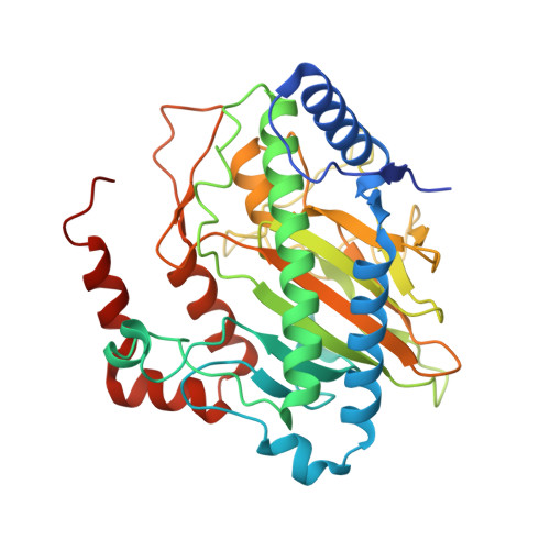
    5LSQ, 5LUN, 5MOF

  • PubMed Abstract: 

    Ethylene is important in industry and biological signaling. In plants, ethylene is produced by oxidation of 1-aminocyclopropane-1-carboxylic acid, as catalyzed by 1-aminocyclopropane-1-carboxylic acid oxidase. Bacteria catalyze ethylene production, but via the four-electron oxidation of 2-oxoglutarate to give ethylene in an arginine-dependent reaction. Crystallographic and biochemical studies on the Pseudomonas syringae ethylene-forming enzyme reveal a branched mechanism. In one branch, an apparently typical 2-oxoglutarate oxygenase reaction to give succinate, carbon dioxide, and sometimes pyrroline-5-carboxylate occurs. Alternatively, Grob-type oxidative fragmentation of a 2-oxoglutarate-derived intermediate occurs to give ethylene and carbon dioxide. Crystallographic and quantum chemical studies reveal that fragmentation to give ethylene is promoted by binding of l-arginine in a nonoxidized conformation and of 2-oxoglutarate in an unprecedented high-energy conformation that favors ethylene, relative to succinate formation.


  • Organizational Affiliation

    Department of Chemistry, University of Oxford, Oxford, OX1 3TA, United Kingdom.


Macromolecules
Find similar proteins by:  (by identity cutoff)  |  3D Structure
Entity ID: 1
MoleculeChains Sequence LengthOrganismDetailsImage
2-oxoglutarate-dependent ethylene/succinate-forming enzyme359Pseudomonas savastanoi pv. phaseolicolaMutation(s): 0 
Gene Names: efe
EC: 1.13.12.19 (PDB Primary Data), 1.14.11.34 (PDB Primary Data)
UniProt
Find proteins for P32021 (Pseudomonas savastanoi pv. phaseolicola)
Explore P32021 
Go to UniProtKB:  P32021
Entity Groups  
Sequence Clusters30% Identity50% Identity70% Identity90% Identity95% Identity100% Identity
UniProt GroupP32021
Sequence Annotations
Expand
  • Reference Sequence
Small Molecules
Ligands 4 Unique
IDChains Name / Formula / InChI Key2D Diagram3D Interactions
AKG
Query on AKG

Download Ideal Coordinates CCD File 
E [auth A],
F [auth A]
2-OXOGLUTARIC ACID
C5 H6 O5
KPGXRSRHYNQIFN-UHFFFAOYSA-N
GOL
Query on GOL

Download Ideal Coordinates CCD File 
G [auth A],
H [auth A]
GLYCEROL
C3 H8 O3
PEDCQBHIVMGVHV-UHFFFAOYSA-N
MN
Query on MN

Download Ideal Coordinates CCD File 
B [auth A],
C [auth A]
MANGANESE (II) ION
Mn
WAEMQWOKJMHJLA-UHFFFAOYSA-N
CL
Query on CL

Download Ideal Coordinates CCD File 
D [auth A]CHLORIDE ION
Cl
VEXZGXHMUGYJMC-UHFFFAOYSA-M
Experimental Data & Validation

Experimental Data

  • Method: X-RAY DIFFRACTION
  • Resolution: 1.45 Å
  • R-Value Free: 0.189 
  • R-Value Work: 0.172 
  • R-Value Observed: 0.173 
  • Space Group: I 2 2 2
Unit Cell:
Length ( Å )Angle ( ˚ )
a = 79.484α = 90
b = 97.85β = 90
c = 98.094γ = 90
Software Package:
Software NamePurpose
Aimlessdata scaling
PHENIXrefinement
PDB_EXTRACTdata extraction
xia2data reduction
PHENIXphasing

Structure Validation

View Full Validation Report


Entry History 

Deposition Data

Revision History  (Full details and data files)

  • Version 1.0: 2017-04-19
    Type: Initial release
  • Version 1.1: 2017-05-03
    Changes: Database references
  • Version 1.2: 2017-05-10
    Changes: Database references
  • Version 2.0: 2024-01-17
    Changes: Atomic model, Data collection, Database references, Derived calculations, Refinement description