5LYC

Cytochrome c in complex with phosphonato-calix[6]arene


Experimental Data Snapshot

  • Method: X-RAY DIFFRACTION
  • Resolution: 1.80 Å
  • R-Value Free: 0.199 
  • R-Value Work: 0.169 
  • R-Value Observed: 0.170 

wwPDB Validation   3D Report Full Report


Ligand Structure Quality Assessment 


This is version 2.0 of the entry. See complete history


Literature

Protein Dimerization on a Phosphonated Calix[6]arene Disc.

Rennie, M.L.Doolan, A.M.Raston, C.L.Crowley, P.B.

(2017) Angew Chem Int Ed Engl 56: 5517-5521

  • DOI: https://doi.org/10.1002/anie.201701500
  • Primary Citation of Related Structures:  
    5LYC

  • PubMed Abstract: 

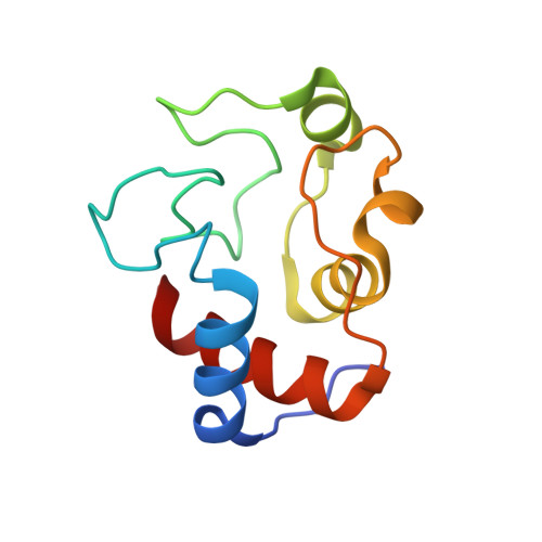
    Complex formation between cationic cytochrome c and the water-soluble, poly-anionic p-phosphonatocalix[6]arene (pclx 6 ) was investigated. A crystal structure (at 1.8 Å resolution) revealed a remarkable dimeric disc of pclx 6 that acts like glue to mediate a symmetric (C 2 ) protein dimer. The calixarene disc has a diameter of about 1.5 nm and masks about 360 Å 2 of protein surface. The key protein-calixarene contacts occur via two linchpin lysines, with additional contacts provided by a small hydrophobic patch. The protein-calixarene supramolecular assemblies were observed in solution by size-exclusion chromatography with multi-angle light scattering and NMR spectroscopy. Using isothermal titration calorimetry and NMR data, an apparent K d in the low micromolar range was determined for the charge-rich protein-calixarene complex. In contrast to p-sulfonatocalix[4]arene, the larger pclx 6 has a single, well-defined binding site that mediates the assembly of cytochrome c in solution.


  • Organizational Affiliation

    School of Chemistry, National University of Ireland Galway, University Road, Galway, Ireland.


Macromolecules
Find similar proteins by:  (by identity cutoff)  |  3D Structure
Entity ID: 1
MoleculeChains Sequence LengthOrganismDetailsImage
Cytochrome c iso-1
A, B
108Saccharomyces cerevisiae S288CMutation(s): 1 
Gene Names: CYC1YJR048WJ1653
UniProt
Find proteins for P00044 (Saccharomyces cerevisiae (strain ATCC 204508 / S288c))
Explore P00044 
Go to UniProtKB:  P00044
Entity Groups  
Sequence Clusters30% Identity50% Identity70% Identity90% Identity95% Identity100% Identity
UniProt GroupP00044
Sequence Annotations
Expand
  • Reference Sequence
Small Molecules
Ligands 3 Unique
IDChains Name / Formula / InChI Key2D Diagram3D Interactions
7AZ
Query on 7AZ

Download Ideal Coordinates CCD File 
D [auth A],
H [auth B]
phosphonato-calix[6]arene
C42 H42 O24 P6
QYHXEDFDCVRPFA-UHFFFAOYSA-N
HEC
Query on HEC

Download Ideal Coordinates CCD File 
C [auth A],
G [auth B]
HEME C
C34 H34 Fe N4 O4
HXQIYSLZKNYNMH-LJNAALQVSA-N
SO4
Query on SO4

Download Ideal Coordinates CCD File 
E [auth A],
F [auth A],
I [auth B],
J [auth B],
K [auth B]
SULFATE ION
O4 S
QAOWNCQODCNURD-UHFFFAOYSA-L
Experimental Data & Validation

Experimental Data

  • Method: X-RAY DIFFRACTION
  • Resolution: 1.80 Å
  • R-Value Free: 0.199 
  • R-Value Work: 0.169 
  • R-Value Observed: 0.170 
  • Space Group: P 43 21 2
Unit Cell:
Length ( Å )Angle ( ˚ )
a = 54.57α = 90
b = 54.57β = 90
c = 208.01γ = 90
Software Package:
Software NamePurpose
MOSFLMdata collection
Aimlessdata scaling
REFMACrefinement
PDB_EXTRACTdata extraction
PHASERphasing

Structure Validation

View Full Validation Report


Ligand Structure Quality Assessment 


Entry History 

Deposition Data

Revision History  (Full details and data files)

  • Version 1.0: 2017-05-10
    Type: Initial release
  • Version 2.0: 2019-09-25
    Changes: Atomic model, Data collection, Derived calculations, Non-polymer description, Structure summary