5LWY

Revised crystal structure of the human adiponectin receptor 2 in complex with a C18 free fatty acid


Experimental Data Snapshot

  • Method: X-RAY DIFFRACTION
  • Resolution: 2.40 Å
  • R-Value Free: 0.224 
  • R-Value Work: 0.198 
  • R-Value Observed: 0.199 

wwPDB Validation   3D Report Full Report


Ligand Structure Quality Assessment 


This is version 1.7 of the entry. See complete history

Re-refinement Note

This entry reflects an alternative modeling of the original data in: 3WXW


Literature

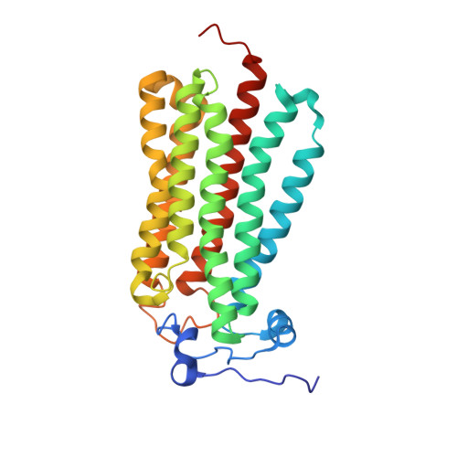
Structural insights into adiponectin receptors suggest ceramidase activity.

Vasiliauskaite-Brooks, I.Sounier, R.Rochaix, P.Bellot, G.Fortier, M.Hoh, F.De Colibus, L.Bechara, C.Saied, E.M.Arenz, C.Leyrat, C.Granier, S.

(2017) Nature 6: 120-123

  • DOI: https://doi.org/10.1038/nature21714
  • Primary Citation of Related Structures:  
    5LWY, 5LX9, 5LXA, 5LXG

  • PubMed Abstract: 

    Adiponectin receptors (ADIPORs) are integral membrane proteins that control glucose and lipid metabolism by mediating, at least in part, a cellular ceramidase activity that catalyses the hydrolysis of ceramide to produce sphingosine and a free fatty acid (FFA). The crystal structures of the two receptor subtypes, ADIPOR1 and ADIPOR2, show a similar overall seven-transmembrane-domain architecture with large unoccupied cavities and a zinc binding site within the seven transmembrane domain. However, the molecular mechanisms by which ADIPORs function are not known. Here we describe the crystal structure of ADIPOR2 bound to a FFA molecule and show that ADIPOR2 possesses intrinsic basal ceramidase activity that is enhanced by adiponectin. We also identify a ceramide binding pose and propose a possible mechanism for the hydrolytic activity of ADIPOR2 using computational approaches. In molecular dynamics simulations, the side chains of residues coordinating the zinc rearrange quickly to promote the nucleophilic attack of a zinc-bound hydroxide ion onto the ceramide amide carbonyl. Furthermore, we present a revised ADIPOR1 crystal structure exhibiting a seven-transmembrane-domain architecture that is clearly distinct from that of ADIPOR2. In this structure, no FFA is observed and the ceramide binding pocket and putative zinc catalytic site are exposed to the inner membrane leaflet. ADIPOR1 also possesses intrinsic ceramidase activity, so we suspect that the two distinct structures may represent key steps in the enzymatic activity of ADIPORs. The ceramidase activity is low, however, and further studies will be required to characterize fully the enzymatic parameters and substrate specificity of ADIPORs. These insights into ADIPOR function will enable the structure-based design of potent modulators of these clinically relevant enzymes.


  • Organizational Affiliation

    Institut de Génomique Fonctionnelle, CNRS UMR-5203 INSERM U1191, University of Montpellier, 34094 Montpellier, France.


Macromolecules
Find similar proteins by:  (by identity cutoff)  |  3D Structure
Entity ID: 1
MoleculeChains Sequence LengthOrganismDetailsImage
Adiponectin receptor protein 2292Homo sapiensMutation(s): 0 
Gene Names: ADIPOR2PAQR2
Membrane Entity: Yes 
UniProt & NIH Common Fund Data Resources
Find proteins for Q86V24 (Homo sapiens)
Explore Q86V24 
Go to UniProtKB:  Q86V24
PHAROS:  Q86V24
GTEx:  ENSG00000006831 
Entity Groups  
Sequence Clusters30% Identity50% Identity70% Identity90% Identity95% Identity100% Identity
UniProt GroupQ86V24
Sequence Annotations
Expand
  • Reference Sequence
Find similar proteins by:  (by identity cutoff)  |  3D Structure
Entity ID: 2
MoleculeChains Sequence LengthOrganismDetailsImage
V REGION HEAVY CHAINB [auth H]119Mus musculusMutation(s): 0 
Entity Groups  
Sequence Clusters30% Identity50% Identity70% Identity90% Identity95% Identity100% Identity
Sequence Annotations
Expand
  • Reference Sequence
Find similar proteins by:  (by identity cutoff)  |  3D Structure
Entity ID: 3
MoleculeChains Sequence LengthOrganismDetailsImage
V REGION LIGHT CHAINC [auth L]107Mus musculusMutation(s): 0 
Entity Groups  
Sequence Clusters30% Identity50% Identity70% Identity90% Identity95% Identity100% Identity
Sequence Annotations
Expand
  • Reference Sequence
Small Molecules
Ligands 4 Unique
IDChains Name / Formula / InChI Key2D Diagram3D Interactions
OLB
Query on OLB

Download Ideal Coordinates CCD File 
E [auth A]
F [auth A]
G [auth A]
H [auth A]
I [auth A]
E [auth A],
F [auth A],
G [auth A],
H [auth A],
I [auth A],
J [auth A],
K [auth A],
L [auth A]
(2S)-2,3-dihydroxypropyl (9Z)-octadec-9-enoate
C21 H40 O4
RZRNAYUHWVFMIP-QJRAZLAKSA-N
OLA
Query on OLA

Download Ideal Coordinates CCD File 
M [auth A]OLEIC ACID
C18 H34 O2
ZQPPMHVWECSIRJ-KTKRTIGZSA-N
GOL
Query on GOL

Download Ideal Coordinates CCD File 
N [auth L]GLYCEROL
C3 H8 O3
PEDCQBHIVMGVHV-UHFFFAOYSA-N
ZN
Query on ZN

Download Ideal Coordinates CCD File 
D [auth A]ZINC ION
Zn
PTFCDOFLOPIGGS-UHFFFAOYSA-N
Experimental Data & Validation

Experimental Data

  • Method: X-RAY DIFFRACTION
  • Resolution: 2.40 Å
  • R-Value Free: 0.224 
  • R-Value Work: 0.198 
  • R-Value Observed: 0.199 
  • Space Group: P 21 2 21
Unit Cell:
Length ( Å )Angle ( ˚ )
a = 74.58α = 90
b = 101.03β = 90
c = 108.63γ = 90
Software Package:
Software NamePurpose
BUSTERrefinement
HKL-2000data reduction
XDSdata scaling
PHASERphasing

Structure Validation

View Full Validation Report


Ligand Structure Quality Assessment 


Entry History & Funding Information

Deposition Data


Funding OrganizationLocationGrant Number
European Research CouncilFrance647687

Revision History  (Full details and data files)

  • Version 1.0: 2017-03-22
    Type: Initial release
  • Version 1.1: 2017-04-05
    Changes: Database references
  • Version 1.2: 2017-04-12
    Changes: Database references
  • Version 1.3: 2017-04-19
    Changes: Atomic model, Structure summary
  • Version 1.4: 2018-02-07
    Changes: Advisory, Author supporting evidence, Database references
  • Version 1.5: 2018-12-05
    Changes: Data collection, Database references, Source and taxonomy, Structure summary
  • Version 1.6: 2019-10-16
    Changes: Data collection
  • Version 1.7: 2024-01-17
    Changes: Advisory, Data collection, Database references, Refinement description