5LLT

Plasmodium falciparum nicotinic acid mononucleotide adenylyltransferase complexed with NaAD


Experimental Data Snapshot

  • Method: X-RAY DIFFRACTION
  • Resolution: 2.20 Å
  • R-Value Free: 0.234 
  • R-Value Work: 0.169 
  • R-Value Observed: 0.172 

wwPDB Validation   3D Report Full Report


Ligand Structure Quality Assessment 


This is version 1.4 of the entry. See complete history


Literature

Structural and Functional Characterization of Plasmodium falciparum Nicotinic Acid Mononucleotide Adenylyltransferase.

Bathke, J.Fritz-Wolf, K.Brandstadter, C.Burkhardt, A.Jortzik, E.Rahlfs, S.Becker, K.

(2016) J Mol Biol 428: 4946-4961

  • DOI: https://doi.org/10.1016/j.jmb.2016.10.023
  • Primary Citation of Related Structures:  
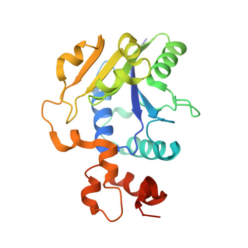
    5LLT, 5LM3

  • PubMed Abstract: 

    Nicotinic acid mononucleotide adenylyltransferase (NaMNAT) is an indispensable enzyme for the synthesis of NAD and NAD phosphate. It catalyzes the adenylylation of nicotinic acid mononucleotide (NaMN) to yield nicotinic acid adenine dinucleotide (NaAD). Since NAD(H) and NAD phosphate(H) are essentially involved in metabolic and redox regulatory reactions, NaMNAT is an attractive drug target in the fight against bacterial and parasitic infections. Notably, NaMNAT of the malaria parasite Plasmodium falciparum possesses only 20% sequence identity with the homologous human enzyme. Here, we present for the first time the two X-ray structures of P. falciparum NaMNAT (PfNaMNAT)-in the product-bound state with NaAD and complexed with an α,β-non-hydrolizable ATP analog-the structures were determined to a resolution of 2.2Å and 2.5Å, respectively. The overall architecture of PfNaMNAT was found to be more similar to its bacterial homologs than its human counterparts although the PPHK motif conserved in bacteria is missing. Furthermore, PfNaMNAT possesses two cysteine residues within the active site that have not been described for any other NaMNATase so far and are likely to be involved in redox regulation of PfNaMNAT activity. Enzymatic studies and surface plasmon resonance data reveal that PfNaMNAT is capable of utilizing NaMN and nicotinamide mononucleotide with a slight preference for NaMN. Surprisingly, a comparison with the active site of Escherichia coli NaMNAT showed very similar architectures, despite different substrate preferences.


  • Organizational Affiliation

    Biochemistry and Molecular Biology, Interdisciplinary Research Center, Justus Liebig University, D-35392 Giessen, Germany.


Macromolecules
Find similar proteins by:  (by identity cutoff)  |  3D Structure
Entity ID: 1
MoleculeChains Sequence LengthOrganismDetailsImage
Nicotinate-nucleotide adenylyltransferase
A, B
213Plasmodium falciparum 3D7Mutation(s): 0 
Gene Names: PF3D7_1327600
EC: 2.7.7.18
UniProt
Find proteins for Q8IE38 (Plasmodium falciparum (isolate 3D7))
Explore Q8IE38 
Go to UniProtKB:  Q8IE38
Entity Groups  
Sequence Clusters30% Identity50% Identity70% Identity90% Identity95% Identity100% Identity
UniProt GroupQ8IE38
Sequence Annotations
Expand
  • Reference Sequence
Small Molecules
Ligands 3 Unique
IDChains Name / Formula / InChI Key2D Diagram3D Interactions
DND
Query on DND

Download Ideal Coordinates CCD File 
C [auth A],
J [auth B]
NICOTINIC ACID ADENINE DINUCLEOTIDE
C21 H27 N6 O15 P2
SENPVEZBRZQVST-HISDBWNOSA-O
PEG
Query on PEG

Download Ideal Coordinates CCD File 
D [auth A],
K [auth B]
DI(HYDROXYETHYL)ETHER
C4 H10 O3
MTHSVFCYNBDYFN-UHFFFAOYSA-N
EDO
Query on EDO

Download Ideal Coordinates CCD File 
E [auth A]
F [auth A]
G [auth A]
H [auth A]
I [auth A]
E [auth A],
F [auth A],
G [auth A],
H [auth A],
I [auth A],
L [auth B],
M [auth B],
N [auth B],
O [auth B]
1,2-ETHANEDIOL
C2 H6 O2
LYCAIKOWRPUZTN-UHFFFAOYSA-N
Experimental Data & Validation

Experimental Data

  • Method: X-RAY DIFFRACTION
  • Resolution: 2.20 Å
  • R-Value Free: 0.234 
  • R-Value Work: 0.169 
  • R-Value Observed: 0.172 
  • Space Group: P 1
Unit Cell:
Length ( Å )Angle ( ˚ )
a = 38.97α = 93.94
b = 52.11β = 90
c = 61.25γ = 109.85
Software Package:
Software NamePurpose
PHENIXrefinement
XDSdata reduction
CNSphasing
Cootmodel building
XSCALEdata scaling

Structure Validation

View Full Validation Report


Ligand Structure Quality Assessment 


Entry History & Funding Information

Deposition Data


Funding OrganizationLocationGrant Number
German Research FoundationGermany(BE1540/23-1 within SPP 1710)

Revision History  (Full details and data files)

  • Version 1.0: 2016-11-30
    Type: Initial release
  • Version 1.1: 2016-12-28
    Changes: Database references
  • Version 1.2: 2017-03-29
    Changes: Database references
  • Version 1.3: 2018-01-31
    Changes: Database references
  • Version 1.4: 2024-01-10
    Changes: Data collection, Database references, Refinement description