5LGB

Crystal structure of murine N1-acetylpolyamine oxidase in complex with MDL72527


Experimental Data Snapshot

  • Method: X-RAY DIFFRACTION
  • Resolution: 1.80 Å
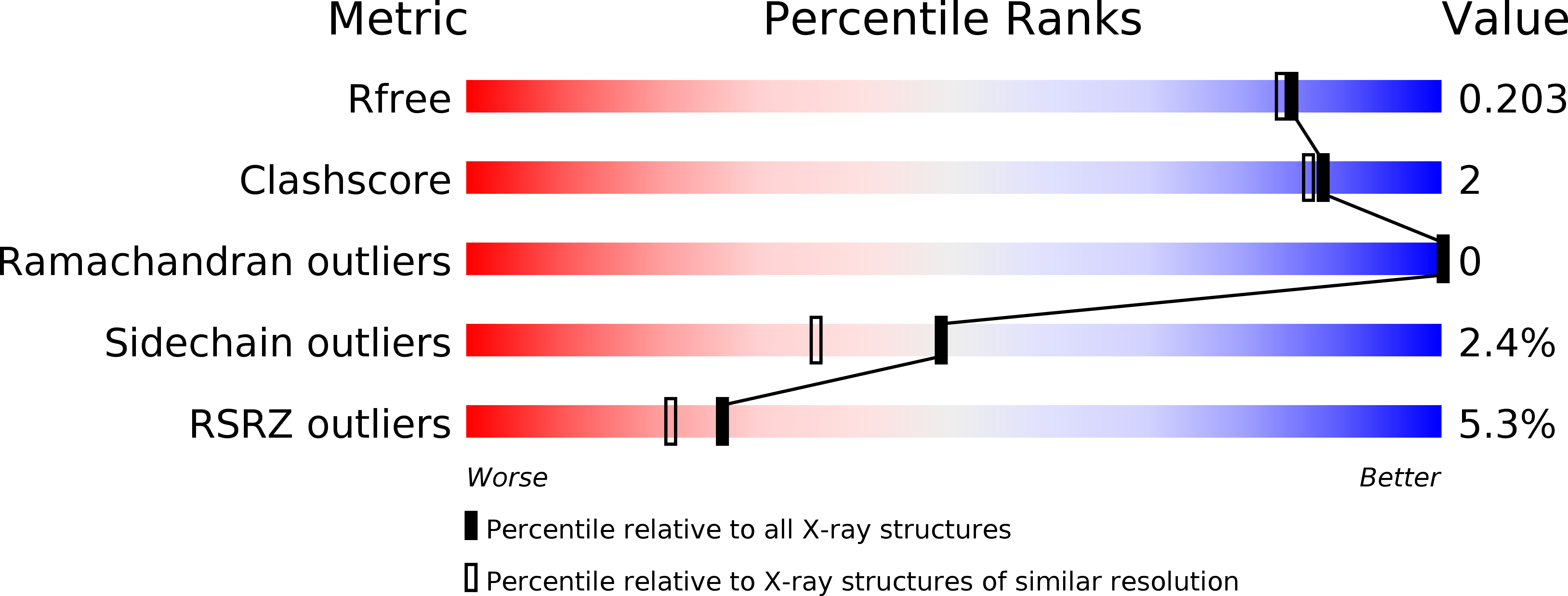
  • R-Value Free: 0.199 
  • R-Value Work: 0.176 
  • R-Value Observed: 0.177 

wwPDB Validation   3D Report Full Report


Ligand Structure Quality Assessment 


This is version 1.1 of the entry. See complete history


Literature

The Structure of Murine N(1)-Acetylspermine Oxidase Reveals Molecular Details of Vertebrate Polyamine Catabolism.

Sjogren, T.Wassvik, C.M.Snijder, A.Aagaard, A.Kumanomidou, T.Barlind, L.Kaminski, T.P.Kashima, A.Yokota, T.Fjellstrom, O.

(2017) Biochemistry 56: 458-467

  • DOI: https://doi.org/10.1021/acs.biochem.6b01140
  • Primary Citation of Related Structures:  
    5LAE, 5LFO, 5LGB, 5MBX

  • PubMed Abstract: 

    N 1 -Acetylspermine oxidase (APAO) catalyzes the conversion of N 1 -acetylspermine or N 1 -acetylspermidine to spermidine or putrescine, respectively, with concomitant formation of N-acetyl-3-aminopropanal and hydrogen peroxide. Here we present the structure of murine APAO in its oxidized holo form and in complex with substrate. The structures provide a basis for understanding molecular details of substrate interaction in vertebrate APAO, highlighting a key role for an asparagine residue in coordinating the N 1 -acetyl group of the substrate. We applied computational methods to the crystal structures to rationalize previous observations with regard to the substrate charge state. The analysis suggests that APAO features an active site ideally suited for binding of charged polyamines. We also reveal the structure of APAO in complex with the irreversible inhibitor MDL72527. In addition to the covalent adduct, a second MDL72527 molecule is bound in the active site. Binding of MDL72527 is accompanied by altered conformations in the APAO backbone. On the basis of structures of APAO, we discuss the potential for development of specific inhibitors.


  • Organizational Affiliation

    Discovery Sciences, Innovative Medicines and Early Development, AstraZeneca , Pepparedsleden 1, SE-431 83 Mölndal, Sweden.


Macromolecules
Find similar proteins by:  (by identity cutoff)  |  3D Structure
Entity ID: 1
MoleculeChains Sequence LengthOrganismDetailsImage
Peroxisomal N(1)-acetyl-spermine/spermidine oxidase,Peroxisomal N(1)-acetyl-spermine/spermidine oxidase497Mus musculusMutation(s): 0 
Gene Names: PaoxPao
EC: 1.5.3.13
UniProt & NIH Common Fund Data Resources
Find proteins for Q8C0L6 (Mus musculus)
Explore Q8C0L6 
Go to UniProtKB:  Q8C0L6
IMPC:  MGI:1916983
Entity Groups  
Sequence Clusters30% Identity50% Identity70% Identity90% Identity95% Identity100% Identity
UniProt GroupQ8C0L6
Sequence Annotations
Expand
  • Reference Sequence
Small Molecules
Ligands 2 Unique
IDChains Name / Formula / InChI Key2D Diagram3D Interactions
6YU
Query on 6YU

Download Ideal Coordinates CCD File 
B [auth A]FAD-MDL72527 adduct
C39 H53 N11 O15 P2
DOENOACRUGDSKX-BYAUSPTGSA-N
MD2
Query on MD2

Download Ideal Coordinates CCD File 
C [auth A]N,N'-BIS(2,3-BUTADIENYL)-1,4-BUTANE-DIAMINE
C12 H20 N2
IKSQCMLJDHRWOA-UHFFFAOYSA-N
Binding Affinity Annotations 
IDSourceBinding Affinity
MD2 BindingDB:  5LGB Ki: 1700 (nM) from 1 assay(s)
Experimental Data & Validation

Experimental Data

  • Method: X-RAY DIFFRACTION
  • Resolution: 1.80 Å
  • R-Value Free: 0.199 
  • R-Value Work: 0.176 
  • R-Value Observed: 0.177 
  • Space Group: P 61
Unit Cell:
Length ( Å )Angle ( ˚ )
a = 121.08α = 90
b = 121.08β = 90
c = 55.03γ = 120
Software Package:
Software NamePurpose
BUSTERrefinement
XDSdata reduction
Aimlessdata scaling
PHASERphasing

Structure Validation

View Full Validation Report


Ligand Structure Quality Assessment 


Entry History 

Deposition Data

Revision History  (Full details and data files)

  • Version 1.0: 2017-08-16
    Type: Initial release
  • Version 1.1: 2024-01-10
    Changes: Data collection, Database references, Refinement description