5L3V

Structure of the crenarchaeal SRP54 GTPase bound to GDP


Experimental Data Snapshot

  • Method: X-RAY DIFFRACTION
  • Resolution: 2.30 Å
  • R-Value Free: 0.222 
  • R-Value Work: 0.176 
  • R-Value Observed: 0.178 

wwPDB Validation   3D Report Full Report


Ligand Structure Quality Assessment 


This is version 1.2 of the entry. See complete history


Literature

Structural Basis for Conserved Regulation and Adaptation of the Signal Recognition Particle Targeting Complex.

Wild, K.Bange, G.Motiejunas, D.Kribelbauer, J.Hendricks, A.Segnitz, B.Wade, R.C.Sinning, I.

(2016) J Mol Biol 428: 2880-2897

  • DOI: https://doi.org/10.1016/j.jmb.2016.05.015
  • Primary Citation of Related Structures:  
    5L3Q, 5L3R, 5L3S, 5L3V, 5L3W

  • PubMed Abstract: 

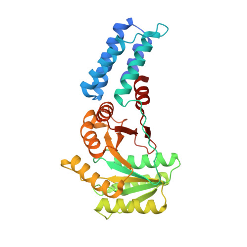
    The signal recognition particle (SRP) is a ribonucleoprotein complex with a key role in targeting and insertion of membrane proteins. The two SRP GTPases, SRP54 (Ffh in bacteria) and FtsY (SRα in eukaryotes), form the core of the targeting complex (TC) regulating the SRP cycle. The architecture of the TC and its stimulation by RNA has been described for the bacterial SRP system while this information is lacking for other domains of life. Here, we present the crystal structures of the GTPase heterodimers of archaeal (Sulfolobus solfataricus), eukaryotic (Homo sapiens), and chloroplast (Arabidopsis thaliana) SRP systems. The comprehensive structural comparison combined with Brownian dynamics simulations of TC formation allows for the description of the general blueprint and of specific adaptations of the quasi-symmetric heterodimer. Our work defines conserved external nucleotide-binding sites for SRP GTPase activation by RNA. Structural analyses of the GDP-bound, post-hydrolysis states reveal a conserved, magnesium-sensitive switch within the I-box. Overall, we provide a general model for SRP cycle regulation by RNA.


  • Organizational Affiliation

    Heidelberg University Biochemistry Center (BZH), INF 328, D-69120 Heidelberg, Germany.


Macromolecules
Find similar proteins by:  (by identity cutoff)  |  3D Structure
Entity ID: 1
MoleculeChains Sequence LengthOrganismDetailsImage
Signal recognition particle 54 kDa protein
A, B
299Saccharolobus solfataricusMutation(s): 0 
Gene Names: srp54SSO0971
UniProt
Find proteins for Q97ZE7 (Saccharolobus solfataricus (strain ATCC 35092 / DSM 1617 / JCM 11322 / P2))
Explore Q97ZE7 
Go to UniProtKB:  Q97ZE7
Entity Groups  
Sequence Clusters30% Identity50% Identity70% Identity90% Identity95% Identity100% Identity
UniProt GroupQ97ZE7
Sequence Annotations
Expand
  • Reference Sequence
Experimental Data & Validation

Experimental Data

  • Method: X-RAY DIFFRACTION
  • Resolution: 2.30 Å
  • R-Value Free: 0.222 
  • R-Value Work: 0.176 
  • R-Value Observed: 0.178 
  • Space Group: P 32 2 1
Unit Cell:
Length ( Å )Angle ( ˚ )
a = 92.168α = 90
b = 92.168β = 90
c = 148.24γ = 120
Software Package:
Software NamePurpose
PHENIXrefinement
iMOSFLMdata reduction
SCALAdata scaling
PHASERphasing

Structure Validation

View Full Validation Report


Ligand Structure Quality Assessment 


Entry History & Funding Information

Deposition Data


Funding OrganizationLocationGrant Number
German Research FoundationGermanyGRK1188

Revision History  (Full details and data files)

  • Version 1.0: 2016-06-08
    Type: Initial release
  • Version 1.1: 2016-07-20
    Changes: Database references
  • Version 1.2: 2024-01-10
    Changes: Author supporting evidence, Data collection, Database references, Refinement description