Engineering a targeted delivery platform using Centyrins.
Goldberg, S.D., Cardoso, R.M., Lin, T., Spinka-Doms, T., Klein, D., Jacobs, S.A., Dudkin, V., Gilliland, G., O'Neil, K.T.(2016) Protein Eng Des Sel 29: 563-572
- PubMed: 27737926
- DOI: https://doi.org/10.1093/protein/gzw054
- Primary Citation of Related Structures:
5L2H - PubMed Abstract:
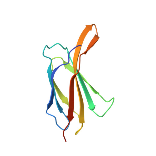
Targeted delivery of therapeutic payloads to specific tissues and cell types is an important component of modern pharmaceutical development. Antibodies or other scaffold proteins can provide the cellular address for delivering a covalently linked therapeutic via specific binding to cell-surface receptors. Optimization of the conjugation site on the targeting protein, linker chemistry and intracellular trafficking pathways can all influence the efficiency of delivery and potency of the drug candidate. In this study, we describe a comprehensive engineering experiment for an EGFR binding Centyrin, a highly stable fibronectin type III (FN3) domain, wherein all possible single-cysteine replacements were evaluated for expression, purification, conjugation efficiency, retention of target binding, biophysical properties and delivery of a cytotoxic small molecule payload. Overall, 26 of the 94 positions were identified as ideal for cysteine modification, conjugation and drug delivery. Conjugation-tolerant positions were mapped onto a crystal structure of the Centyrin, providing a structural context for interpretation of the mutagenesis experiment and providing a foundation for a Centyrin-targeted delivery platform.
Organizational Affiliation:
Janssen Research and Development, L.L.C., 1400 McKean Road, Spring House, PA 19477, USA.