5KQA

Crystal structure of buckwheat glutaredoxin-glutathione complex


Experimental Data Snapshot

  • Method: X-RAY DIFFRACTION
  • Resolution: 2.05 Å
  • R-Value Free: 0.216 
  • R-Value Work: 0.163 
  • R-Value Observed: 0.168 

wwPDB Validation   3D Report Full Report


Ligand Structure Quality Assessment 


This is version 1.2 of the entry. See complete history


Literature

Structural insights into the binding of buckwheat glutaredoxin with GSH and regulation of its catalytic activity

Zhang, X.Wang, W.Li, C.Zhao, Y.Yuan, H.Tan, X.Wu, L.Wang, Z.Wang, H.

(2017) J Inorg Biochem 173: 21-27

  • DOI: https://doi.org/10.1016/j.jinorgbio.2017.04.019
  • Primary Citation of Related Structures:  
    5GTX, 5KQA

  • PubMed Abstract: 

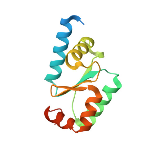
    Glutaredoxins (Grxs) are ubiquitous thioltransferases and members of the thioredoxin (Trx) fold superfamily. They have multiple functions in cells including oxidative stress responses and cell signaling. A novel glutaredoxin from buckwheat (rbGrx) with higher catalytic activity was identified, cloned, and purified. The structures of glutathionylated rbGrx and an rbGrx mutant, in which cysteine 39 was mutated to alanine, were solved by x-ray diffraction at a resolution of 2.05Å and 2.29Å, respectively. In rbGrx, GSH (glutathione) is bound at the conserved GSH-binding site, and its structure shows that it has the potential to function as a scaffold protein for the assembly and delivery of GSH. The crystal structure shows that GSH does not bind to the C39A rbGrx mutant, and the C39A mutant had no catalytic activity, indicating that C39 is a key residue that is involved in both the binding of rbGrx to GSH and the regulation of its catalytic activity. The model showing the binding of GSH with rbGrx provides a basis for understanding its molecular function and its potential future applications in medicinal food science.


  • Organizational Affiliation

    College of Life Science, Shanxi University, Shanxi, Taiyuan 030006, China.


Macromolecules
Find similar proteins by:  (by identity cutoff)  |  3D Structure
Entity ID: 1
MoleculeChains Sequence LengthOrganismDetailsImage
Glutaredoxin-glutathione complex132PolygonaceaeMutation(s): 0 
UniProt
Find proteins for A0A1W6I4R7 (Fagopyrum tataricum)
Explore A0A1W6I4R7 
Go to UniProtKB:  A0A1W6I4R7
Entity Groups  
Sequence Clusters30% Identity50% Identity70% Identity90% Identity95% Identity100% Identity
UniProt GroupA0A1W6I4R7
Sequence Annotations
Expand
  • Reference Sequence
Small Molecules
Ligands 1 Unique
IDChains Name / Formula / InChI Key2D Diagram3D Interactions
GSH
Query on GSH

Download Ideal Coordinates CCD File 
B [auth A]GLUTATHIONE
C10 H17 N3 O6 S
RWSXRVCMGQZWBV-WDSKDSINSA-N
Experimental Data & Validation

Experimental Data

  • Method: X-RAY DIFFRACTION
  • Resolution: 2.05 Å
  • R-Value Free: 0.216 
  • R-Value Work: 0.163 
  • R-Value Observed: 0.168 
  • Space Group: P 21 21 2
Unit Cell:
Length ( Å )Angle ( ˚ )
a = 43.011α = 90
b = 92.299β = 90
c = 28.133γ = 90
Software Package:
Software NamePurpose
PHENIXrefinement
HKL-3000data reduction
HKL-3000data scaling
PHASERphasing

Structure Validation

View Full Validation Report


Ligand Structure Quality Assessment 


Entry History & Funding Information

Deposition Data


Funding OrganizationLocationGrant Number
National Natural Science Foundation of ChinaChina2014DFA30270
National Natural Science Foundation of ChinaChina21543003
Hundred Talent Plans and Research Projects of Shanxi ProvinceChina2015081049

Revision History  (Full details and data files)

  • Version 1.0: 2017-07-05
    Type: Initial release
  • Version 1.1: 2017-09-27
    Changes: Data collection
  • Version 1.2: 2023-11-08
    Changes: Data collection, Database references, Refinement description