5KOR

Arabidopsis thaliana fucosyltransferase 1 (FUT1) in complex with GDP and a xylo-oligossacharide


Experimental Data Snapshot

  • Method: X-RAY DIFFRACTION
  • Resolution: 2.20 Å
  • R-Value Free: 0.215 
  • R-Value Work: 0.179 
  • R-Value Observed: 0.181 

wwPDB Validation   3D Report Full Report


Ligand Structure Quality Assessment 


This is version 2.1 of the entry. See complete history


Literature

Structure of Arabidopsis thaliana FUT1 Reveals a Variant of the GT-B Class Fold and Provides Insight into Xyloglucan Fucosylation.

Rocha, J.Ciceron, F.de Sanctis, D.Lelimousin, M.Chazalet, V.Lerouxel, O.Breton, C.

(2016) Plant Cell 28: 2352-2364

  • DOI: https://doi.org/10.1105/tpc.16.00519
  • Primary Citation of Related Structures:  
    5KOP, 5KOR

  • PubMed Abstract: 

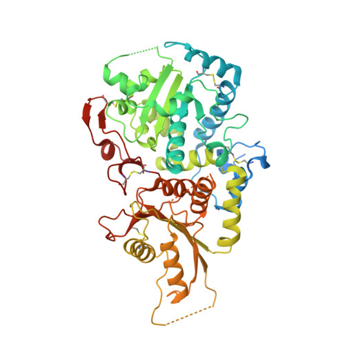
    The plant cell wall is a complex and dynamic network made mostly of cellulose, hemicelluloses, and pectins. Xyloglucan, the major hemicellulosic component in Arabidopsis thaliana, is biosynthesized in the Golgi apparatus by a series of glycan synthases and glycosyltransferases before export to the wall. A better understanding of the xyloglucan biosynthetic machinery will give clues toward engineering plants with improved wall properties or designing novel xyloglucan-based biomaterials. The xyloglucan-specific α2-fucosyltransferase FUT1 catalyzes the transfer of fucose from GDP-fucose to terminal galactosyl residues on xyloglucan side chains. Here, we present crystal structures of Arabidopsis FUT1 in its apoform and in a ternary complex with GDP and a xylo-oligosaccharide acceptor (named XLLG). Although FUT1 is clearly a member of the large GT-B fold family, like other fucosyltransferases of known structures, it contains a variant of the GT-B fold. In particular, it includes an extra C-terminal region that is part of the acceptor binding site. Our crystal structures support previous findings that FUT1 behaves as a functional dimer. Mutational studies and structure comparison with other fucosyltransferases suggest that FUT1 uses a S N 2-like reaction mechanism similar to that of protein-O-fucosyltransferase 2. Thus, our results provide new insights into the mechanism of xyloglucan fucosylation in the Golgi.


  • Organizational Affiliation

    Université Grenoble Alpes, 38400 Grenoble, France.


Macromolecules
Find similar proteins by:  (by identity cutoff)  |  3D Structure
Entity ID: 1
MoleculeChains Sequence LengthOrganismDetailsImage
Galactoside 2-alpha-L-fucosyltransferase
A, B, C, D
521Arabidopsis thalianaMutation(s): 0 
Gene Names: FUT1FT1MUR2At2g03220T18E12.11
EC: 2.4.1.69
UniProt
Find proteins for Q9SWH5 (Arabidopsis thaliana)
Explore Q9SWH5 
Go to UniProtKB:  Q9SWH5
Entity Groups  
Sequence Clusters30% Identity50% Identity70% Identity90% Identity95% Identity100% Identity
UniProt GroupQ9SWH5
Sequence Annotations
Expand
  • Reference Sequence
Oligosaccharides

Help

Entity ID: 2
MoleculeChains Length2D Diagram Glycosylation3D Interactions
alpha-D-xylopyranose-(1-6)-beta-D-glucopyranose-(1-4)-[beta-D-galactopyranose-(1-2)-alpha-D-xylopyranose-(1-6)]beta-D-glucopyranose-(1-4)-[beta-D-galactopyranose-(1-2)-alpha-D-xylopyranose-(1-6)]beta-D-glucopyranose-(1-4)-beta-D-glucopyranose
E, F
9N/A
Glycosylation Resources
GlyTouCan:  G06184LI
GlyCosmos:  G06184LI
Small Molecules
Ligands 3 Unique
IDChains Name / Formula / InChI Key2D Diagram3D Interactions
GDP
Query on GDP

Download Ideal Coordinates CCD File 
G [auth A],
K [auth B],
O [auth C],
W [auth D]
GUANOSINE-5'-DIPHOSPHATE
C10 H15 N5 O11 P2
QGWNDRXFNXRZMB-UUOKFMHZSA-N
EDO
Query on EDO

Download Ideal Coordinates CCD File 
H [auth A]
I [auth A]
J [auth A]
L [auth B]
M [auth B]
H [auth A],
I [auth A],
J [auth A],
L [auth B],
M [auth B],
P [auth C],
Q [auth C],
R [auth C],
S [auth C],
T [auth C],
U [auth C],
X [auth D],
Y [auth D],
Z [auth D]
1,2-ETHANEDIOL
C2 H6 O2
LYCAIKOWRPUZTN-UHFFFAOYSA-N
CL
Query on CL

Download Ideal Coordinates CCD File 
AA [auth D],
N [auth B],
V [auth C]
CHLORIDE ION
Cl
VEXZGXHMUGYJMC-UHFFFAOYSA-M
Experimental Data & Validation

Experimental Data

  • Method: X-RAY DIFFRACTION
  • Resolution: 2.20 Å
  • R-Value Free: 0.215 
  • R-Value Work: 0.179 
  • R-Value Observed: 0.181 
  • Space Group: P 1 21 1
Unit Cell:
Length ( Å )Angle ( ˚ )
a = 87.975α = 90
b = 85.806β = 96.19
c = 150.723γ = 90
Software Package:
Software NamePurpose
PHENIXrefinement
XDSdata reduction
Aimlessdata scaling
PHASERphasing

Structure Validation

View Full Validation Report


Ligand Structure Quality Assessment 


Entry History & Funding Information

Deposition Data


Funding OrganizationLocationGrant Number
Univ. Grenoble AlpesFrance--

Revision History  (Full details and data files)

  • Version 1.0: 2016-10-12
    Type: Initial release
  • Version 1.1: 2016-11-23
    Changes: Database references
  • Version 1.2: 2018-10-10
    Changes: Data collection, Database references, Source and taxonomy, Structure summary
  • Version 2.0: 2020-07-29
    Type: Remediation
    Reason: Carbohydrate remediation
    Changes: Atomic model, Data collection, Derived calculations, Structure summary
  • Version 2.1: 2024-01-10
    Changes: Data collection, Database references, Derived calculations, Refinement description, Structure summary