


Crystal structure of Mycobacterium tuberculosis hypoxanthine guanine phosphoribosyltransferase in complex with pyrrolidine nucleoside phosphonate
Eng, W.S., Rejman, D., Keough, D.T., Guddat, L.W.To be published.
Experimental Data Snapshot
Starting Model: experimental
View more details
Entity ID: 1 | |||||
|---|---|---|---|---|---|
| Molecule | Chains | Sequence Length | Organism | Details | Image |
| Hypoxanthine-guanine phosphoribosyltransferase | 207 | Mycobacterium tuberculosis H37Rv | Mutation(s): 0 Gene Names: hpt, hprT, Rv3624c, MTCY15C10.28 EC: 2.4.2.8 | 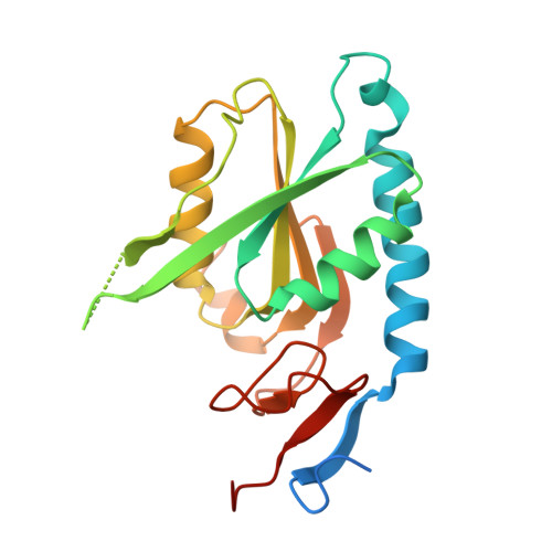 | |
UniProt | |||||
Find proteins for P9WHQ9 (Mycobacterium tuberculosis (strain ATCC 25618 / H37Rv)) Explore P9WHQ9 Go to UniProtKB: P9WHQ9 | |||||
Entity Groups | |||||
| Sequence Clusters | 30% Identity50% Identity70% Identity90% Identity95% Identity100% Identity | ||||
| UniProt Group | P9WHQ9 | ||||
Sequence AnnotationsExpand | |||||
| |||||
| Ligands 3 Unique | |||||
|---|---|---|---|---|---|
| ID | Chains | Name / Formula / InChI Key | 2D Diagram | 3D Interactions | |
| 6W7 Query on 6W7 | E [auth A], I [auth B], M [auth C], Q [auth D] | [(3~{S},4~{R})-4-(6-oxidanylidene-1~{H}-purin-9-yl)pyrrolidin-3-yl]oxymethylphosphonic acid C10 H14 N5 O5 P YFBRPISOXMPUHB-RQJHMYQMSA-N |  | ||
| POP Query on POP | H [auth A], L [auth B], P [auth C], T [auth D] | PYROPHOSPHATE 2- H2 O7 P2 XPPKVPWEQAFLFU-UHFFFAOYSA-L |  | ||
| MG Query on MG | F [auth A] G [auth A] J [auth B] K [auth B] N [auth C] | MAGNESIUM ION Mg JLVVSXFLKOJNIY-UHFFFAOYSA-N |  | ||
| Length ( Å ) | Angle ( ˚ ) |
|---|---|
| a = 55.264 | α = 90 |
| b = 85.678 | β = 90 |
| c = 154.586 | γ = 90 |
| Software Name | Purpose |
|---|---|
| PHENIX | refinement |
| PDB_EXTRACT | data extraction |
| XDS | data reduction |
| SCALA | data scaling |
| PHASER | phasing |
RCSB PDB (citation) is hosted by
RCSB PDB is a member of the

