5KIJ

Crystal structure of the class I human endoplasmic reticulum 1,2-alpha-mannosidase and Man9GlcNAc2-PA complex

  • Classification: HYDROLASE
  • Organism(s): Homo sapiens
  • Expression System: Komagataella pastoris
  • Mutation(s): No 

  • Deposited: 2016-06-16 Released: 2017-05-10 
  • Deposition Author(s): Karaveg, K., Xiang, Y., Moremen, K.W.
  • Funding Organization(s): National Institutes of Health/National Institute of General Medical Sciences (NIH/NIGMS), National Institutes of Health/National Institute of Diabetes and Digestive and Kidney Disease (NIH/NIDDK)

Experimental Data Snapshot

  • Method: X-RAY DIFFRACTION
  • Resolution: 1.65 Å
  • R-Value Free: 0.222 
  • R-Value Work: 0.182 
  • R-Value Observed: 0.184 

wwPDB Validation   3D Report Full Report


Ligand Structure Quality Assessment 


This is version 2.1 of the entry. See complete history


Literature

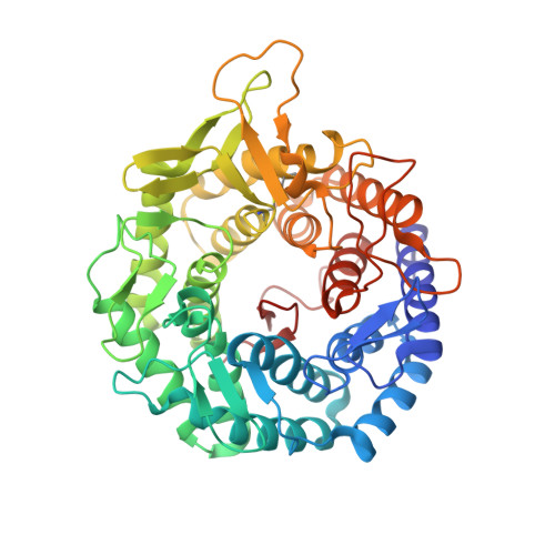
Substrate recognition and catalysis by GH47 alpha-mannosidases involved in Asn-linked glycan maturation in the mammalian secretory pathway.

Xiang, Y.Karaveg, K.Moremen, K.W.

(2016) Proc Natl Acad Sci U S A 113: E7890-E7899

  • DOI: https://doi.org/10.1073/pnas.1611213113
  • Primary Citation of Related Structures:  
    5KIJ, 5KK7, 5KKB

  • PubMed Abstract: 

    Maturation of Asn-linked oligosaccharides in the eukaryotic secretory pathway requires the trimming of nascent glycan chains to remove all glucose and several mannose residues before extension into complex-type structures on the cell surface and secreted glycoproteins. Multiple glycoside hydrolase family 47 (GH47) α-mannosidases, including endoplasmic reticulum (ER) α-mannosidase I (ERManI) and Golgi α-mannosidase IA (GMIA), are responsible for cleavage of terminal α1,2-linked mannose residues to produce uniquely trimmed oligomannose isomers that are necessary for ER glycoprotein quality control and glycan maturation. ERManI and GMIA have similar catalytic domain structures, but each enzyme cleaves distinct residues from tribranched oligomannose glycan substrates. The structural basis for branch-specific cleavage by ERManI and GMIA was explored by replacing an essential enzyme-bound Ca 2+ ion with a lanthanum (La 3+ ) ion. This ion swap led to enzyme inactivation while retaining high-affinity substrate interactions. Cocrystallization of La 3+ -bound enzymes with Man 9 GlcNAc 2 substrate analogs revealed enzyme-substrate complexes with distinct modes of glycan branch insertion into the respective enzyme active-site clefts. Both enzymes had glycan interactions that extended across the entire glycan structure, but each enzyme engaged a different glycan branch and used different sets of glycan interactions. Additional mutagenesis and time-course studies of glycan cleavage probed the structural basis of enzyme specificity. The results provide insights into the enzyme catalytic mechanisms and reveal structural snapshots of the sequential glycan cleavage events. The data also indicate that full steric access to glycan substrates determines the efficiency of mannose-trimming reactions that control the conversion to complex-type structures in mammalian cells.


  • Organizational Affiliation

    Complex Carbohydrate Research Center, University of Georgia, Athens, GA 30602.


Macromolecules
Find similar proteins by:  (by identity cutoff)  |  3D Structure
Entity ID: 1
MoleculeChains Sequence LengthOrganismDetailsImage
Endoplasmic reticulum mannosyl-oligosaccharide 1,2-alpha-mannosidase452Homo sapiensMutation(s): 0 
Gene Names: MAN1B1UNQ747/PRO1477
EC: 3.2.1.113
UniProt & NIH Common Fund Data Resources
Find proteins for Q9UKM7 (Homo sapiens)
Explore Q9UKM7 
Go to UniProtKB:  Q9UKM7
PHAROS:  Q9UKM7
GTEx:  ENSG00000177239 
Entity Groups  
Sequence Clusters30% Identity50% Identity70% Identity90% Identity95% Identity100% Identity
UniProt GroupQ9UKM7
Sequence Annotations
Expand
  • Reference Sequence
Oligosaccharides

Help

Entity ID: 2
MoleculeChains Length2D Diagram Glycosylation3D Interactions
alpha-D-mannopyranose-(1-2)-alpha-D-mannopyranose-(1-3)-[alpha-D-mannopyranose-(1-2)-alpha-D-mannopyranose-(1-6)]alpha-D-mannopyranose-(1-6)-[alpha-D-mannopyranose-(1-2)-alpha-D-mannopyranose-(1-3)]beta-D-mannopyranose-(1-4)-2-acetamido-2-deoxy-beta-D-glucopyranose
B
9N/A
Glycosylation Resources
GlyTouCan:  G51034LA
GlyCosmos:  G51034LA
GlyGen:  G51034LA
Small Molecules
Ligands 4 Unique
IDChains Name / Formula / InChI Key2D Diagram3D Interactions
1PS
Query on 1PS

Download Ideal Coordinates CCD File 
H [auth A]3-PYRIDINIUM-1-YLPROPANE-1-SULFONATE
C8 H11 N O3 S
REEBJQTUIJTGAL-UHFFFAOYSA-N
LA
Query on LA

Download Ideal Coordinates CCD File 
C [auth A]LANTHANUM (III) ION
La
CZMAIROVPAYCMU-UHFFFAOYSA-N
SO4
Query on SO4

Download Ideal Coordinates CCD File 
D [auth A],
E [auth A]
SULFATE ION
O4 S
QAOWNCQODCNURD-UHFFFAOYSA-L
BU1
Query on BU1

Download Ideal Coordinates CCD File 
F [auth A],
G [auth A],
I [auth A],
J [auth A]
1,4-BUTANEDIOL
C4 H10 O2
WERYXYBDKMZEQL-UHFFFAOYSA-N
Experimental Data & Validation

Experimental Data

  • Method: X-RAY DIFFRACTION
  • Resolution: 1.65 Å
  • R-Value Free: 0.222 
  • R-Value Work: 0.182 
  • R-Value Observed: 0.184 
  • Space Group: P 1
Unit Cell:
Length ( Å )Angle ( ˚ )
a = 50.625α = 89.65
b = 53.83β = 63.6
c = 56.118γ = 62.7
Software Package:
Software NamePurpose
PHENIXrefinement
HKL-2000data reduction
HKL-2000data scaling
PHASERphasing

Structure Validation

View Full Validation Report


Ligand Structure Quality Assessment 


Entry History & Funding Information

Deposition Data


Funding OrganizationLocationGrant Number
National Institutes of Health/National Institute of General Medical Sciences (NIH/NIGMS)United StatesR01GM047533
National Institutes of Health/National Institute of Diabetes and Digestive and Kidney Disease (NIH/NIDDK)United StatesR01DK075322
National Institutes of Health/National Institute of General Medical Sciences (NIH/NIGMS)United StatesP41GM103390
National Institutes of Health/National Institute of General Medical Sciences (NIH/NIGMS)United StatesP01GM107012

Revision History  (Full details and data files)

  • Version 1.0: 2017-05-10
    Type: Initial release
  • Version 1.1: 2017-09-06
    Changes: Author supporting evidence
  • Version 1.2: 2019-12-25
    Changes: Author supporting evidence
  • Version 2.0: 2020-07-29
    Type: Remediation
    Reason: Carbohydrate remediation
    Changes: Advisory, Atomic model, Data collection, Derived calculations, Structure summary
  • Version 2.1: 2023-09-27
    Changes: Data collection, Database references, Derived calculations, Refinement description, Structure summary