5KBJ

Structure of Rep-DNA complex


Experimental Data Snapshot

  • Method: X-RAY DIFFRACTION
  • Resolution: 3.09 Å
  • R-Value Free: 0.277 
  • R-Value Work: 0.225 
  • R-Value Observed: 0.231 

wwPDB Validation   3D Report Full Report


This is version 1.2 of the entry. See complete history


Literature

Mechanism of staphylococcal multiresistance plasmid replication origin assembly by the RepA protein.

Schumacher, M.A.Tonthat, N.K.Kwong, S.M.Chinnam, N.B.Liu, M.A.Skurray, R.A.Firth, N.

(2014) Proc Natl Acad Sci U S A 111: 9121-9126

  • DOI: https://doi.org/10.1073/pnas.1406065111
  • Primary Citation of Related Structures:  
    4PQK, 4PQL, 4PT7, 4PTA, 5KBJ

  • PubMed Abstract: 

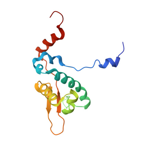
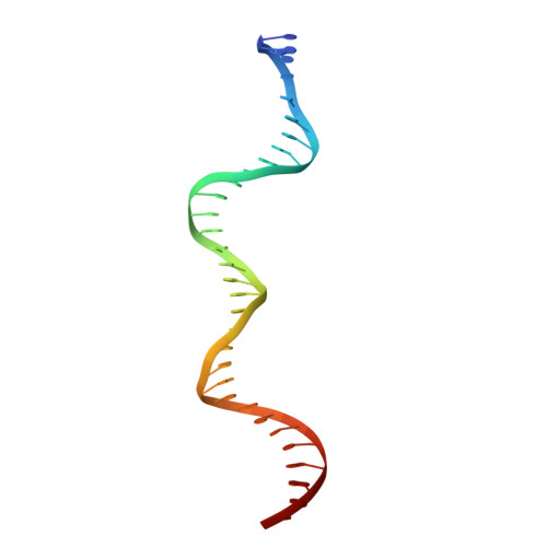
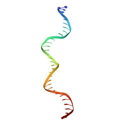
    The staphylococcal multiresistance plasmids are key contributors to the alarming rise in bacterial multidrug resistance. A conserved replication initiator, RepA, encoded on these plasmids is essential for their propagation. RepA proteins consist of flexibly linked N-terminal (NTD) and C-terminal (CTD) domains. Despite their essential role in replication, the molecular basis for RepA function is unknown. Here we describe a complete structural and functional dissection of RepA proteins. Unexpectedly, both the RepA NTD and CTD show similarity to the corresponding domains of the bacterial primosome protein, DnaD. Although the RepA and DnaD NTD both contain winged helix-turn-helices, the DnaD NTD self-assembles into large scaffolds whereas the tetrameric RepA NTD binds DNA iterons using a newly described DNA binding mode. Strikingly, structural and atomic force microscopy data reveal that the NTD tetramer mediates DNA bridging, suggesting a molecular mechanism for origin handcuffing. Finally, data show that the RepA CTD interacts with the host DnaG primase, which binds the replicative helicase. Thus, these combined data reveal the molecular mechanism by which RepA mediates the specific replicon assembly of staphylococcal multiresistant plasmids.


  • Organizational Affiliation

    Department of Biochemistry, Duke University Medical Center, Durham, NC 27710; and maria.schumacher@duke.edu.


Macromolecules

Find similar proteins by:  (by identity cutoff)  |  3D Structure
Entity ID: 1
MoleculeChains Sequence LengthOrganismDetailsImage
Replication initiator A, N-terminal132Staphylococcus aureusMutation(s): 0 
Gene Names: SAP042A_013SAP058A_012SAP071A_014
UniProt
Find proteins for D2JDC3 (Staphylococcus aureus)
Explore D2JDC3 
Go to UniProtKB:  D2JDC3
Entity Groups  
Sequence Clusters30% Identity50% Identity70% Identity90% Identity95% Identity100% Identity
UniProt GroupD2JDC3
Sequence Annotations
Expand
  • Reference Sequence
Find similar nucleic acids by:  (by identity cutoff)  |  3D Structure
Entity ID: 2
MoleculeChains LengthOrganismImage
DNA (32-MER)I [auth R]32synthetic construct
Sequence Annotations
Expand
  • Reference Sequence
Find similar nucleic acids by:  (by identity cutoff)  |  3D Structure
Entity ID: 3
MoleculeChains LengthOrganismImage
DNA (32-MER)J [auth W]32synthetic construct
Sequence Annotations
Expand
  • Reference Sequence
Experimental Data & Validation

Experimental Data

  • Method: X-RAY DIFFRACTION
  • Resolution: 3.09 Å
  • R-Value Free: 0.277 
  • R-Value Work: 0.225 
  • R-Value Observed: 0.231 
  • Space Group: P 1
Unit Cell:
Length ( Å )Angle ( ˚ )
a = 59.2α = 80.8
b = 77.5β = 75.5
c = 100.7γ = 71.9
Software Package:
Software NamePurpose
PHENIXrefinement
PDB_EXTRACTdata extraction
MOSFLMdata reduction
SCALAdata scaling
MOLREPphasing

Structure Validation

View Full Validation Report


Entry History 

Deposition Data

Revision History  (Full details and data files)

  • Version 1.0: 2016-06-29
    Type: Initial release
  • Version 1.1: 2017-11-22
    Changes: Derived calculations, Refinement description
  • Version 1.2: 2023-09-27
    Changes: Data collection, Database references, Refinement description