5K93

PapD wild-type chaperone

  • Classification: CHAPERONE
  • Organism(s): Escherichia coli
  • Expression System: Escherichia coli
  • Mutation(s): No 

  • Deposited: 2016-05-31 Released: 2016-07-20 
  • Deposition Author(s): Sarowar, S., Li, H.
  • Funding Organization(s): National Institutes of Health/National Institute of General Medical Sciences (NIH/NIGMS)

Experimental Data Snapshot

  • Method: X-RAY DIFFRACTION
  • Resolution: 2.70 Å
  • R-Value Free: 0.279 
  • R-Value Work: 0.220 
  • R-Value Observed: 0.223 

wwPDB Validation   3D Report Full Report


This is version 1.4 of the entry. See complete history


Literature

The Escherichia coli P and Type 1 Pilus Assembly Chaperones PapD and FimC Are Monomeric in Solution.

Sarowar, S.Hu, O.J.Werneburg, G.T.Thanassi, D.G.Li, H.

(2016) J Bacteriol 198: 2360-2369

  • DOI: https://doi.org/10.1128/JB.00366-16
  • Primary Citation of Related Structures:  
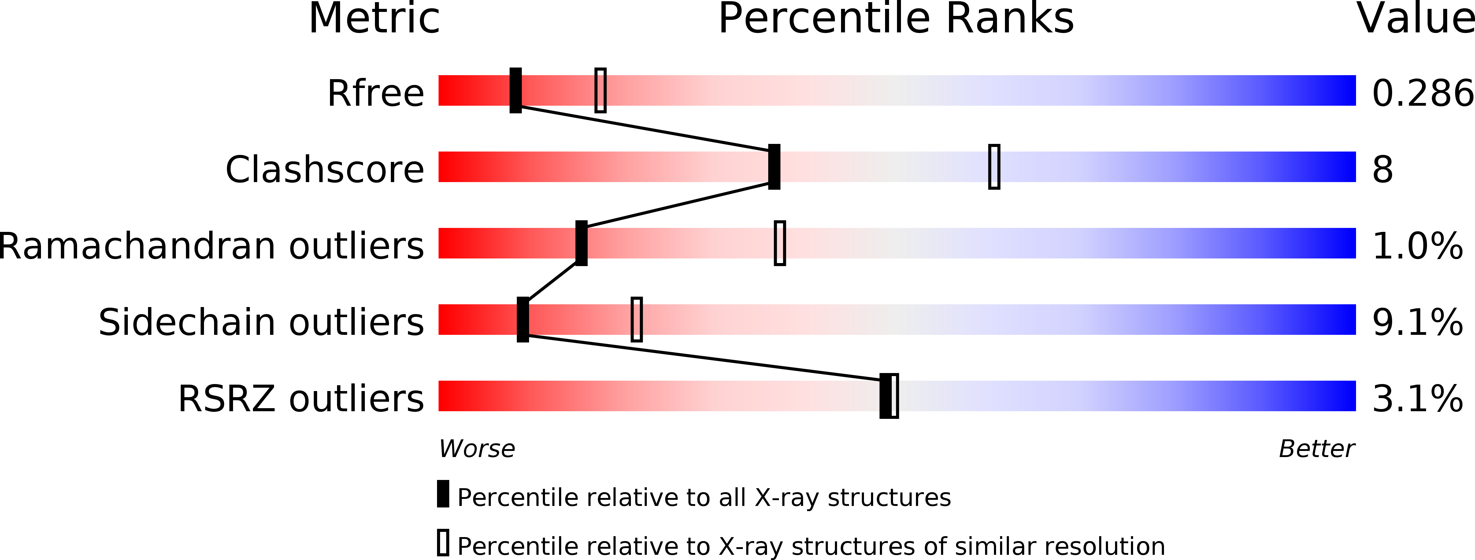
    5K93

  • PubMed Abstract: 

    The chaperone/usher pathway is used by Gram-negative bacteria to assemble adhesive surface structures known as pili or fimbriae. Uropathogenic strains of Escherichia coli use this pathway to assemble P and type 1 pili, which facilitate colonization of the kidney and bladder, respectively. Pilus assembly requires a periplasmic chaperone and outer membrane protein termed the usher. The chaperone allows folding of pilus subunits and escorts the subunits to the usher for polymerization into pili and secretion to the cell surface. Based on previous structures of mutant versions of the P pilus chaperone PapD, it was suggested that the chaperone dimerizes in the periplasm as a self-capping mechanism. Such dimerization is counterintuitive because the chaperone G1 strand, important for chaperone-subunit interaction, is buried at the dimer interface. Here, we show that the wild-type PapD chaperone also forms a dimer in the crystal lattice; however, the dimer interface is different from the previously solved structures. In contrast to the crystal structures, we found that both PapD and the type 1 pilus chaperone, FimC, are monomeric in solution. Our findings indicate that pilus chaperones do not sequester their G1 β-strand by forming a dimer. Instead, the chaperones may expose their G1 strand for facile interaction with pilus subunits. We also found that the type 1 pilus adhesin, FimH, is flexible in solution while in complex with its chaperone, whereas the P pilus adhesin, PapGII, is rigid. Our study clarifies a crucial step in pilus biogenesis and reveals pilus-specific differences that may relate to biological function.


  • Organizational Affiliation

    Department of Biochemistry and Cell Biology, Stony Brook University, Stonybrook, New York, USA Biology Department, Brookhaven National Laboratory, Stonybrook, New York, USA.


Macromolecules
Find similar proteins by:  (by identity cutoff)  |  3D Structure
Entity ID: 1
MoleculeChains Sequence LengthOrganismDetailsImage
Chaperone protein PapD
A, B
222Escherichia coliMutation(s): 0 
Gene Names: papD
UniProt
Find proteins for P15319 (Escherichia coli)
Explore P15319 
Go to UniProtKB:  P15319
Entity Groups  
Sequence Clusters30% Identity50% Identity70% Identity90% Identity95% Identity100% Identity
UniProt GroupP15319
Sequence Annotations
Expand
  • Reference Sequence
Experimental Data & Validation

Experimental Data

  • Method: X-RAY DIFFRACTION
  • Resolution: 2.70 Å
  • R-Value Free: 0.279 
  • R-Value Work: 0.220 
  • R-Value Observed: 0.223 
  • Space Group: C 1 2 1
Unit Cell:
Length ( Å )Angle ( ˚ )
a = 83.129α = 90
b = 70.989β = 102.05
c = 93.05γ = 90
Software Package:
Software NamePurpose
REFMACrefinement
HKL-2000data reduction
HKL-2000data scaling
PHENIXphasing

Structure Validation

View Full Validation Report


Entry History & Funding Information

Deposition Data


Funding OrganizationLocationGrant Number
National Institutes of Health/National Institute of General Medical Sciences (NIH/NIGMS)United StatesT32 GM008468
National Institutes of Health/National Institute of General Medical Sciences (NIH/NIGMS)United StatesR01 GM062987

Revision History  (Full details and data files)

  • Version 1.0: 2016-07-20
    Type: Initial release
  • Version 1.1: 2016-08-24
    Changes: Database references
  • Version 1.2: 2017-09-06
    Changes: Data collection, Database references, Derived calculations
  • Version 1.3: 2017-09-20
    Changes: Author supporting evidence
  • Version 1.4: 2019-12-25
    Changes: Author supporting evidence